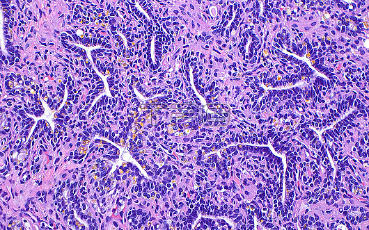
Light micrograph of the epithelium of a seminal vesicle. The seminal vesicle epithelium is formed of glands with irregular lumens. Yellow pigment, which is also characteristic of seminal vesicle epithelium, can also be seen in this image. Haematoxylin and eosin stained tissue section. Magnification: 200x when printed at 10 centimetres.
| px | px | dpi | = | cm | x | cm | = | MB |
Details
Creative#:
TOP30048852
Source:
達志影像
Authorization Type:
RM
Release Information:
須由TPG 完整授權
Model Release:
N/A
Property Release:
N/A
Right to Privacy:
No
Same folder images:
pathologypathologicalmedicineanatomicalpathologypathologicalseminalvesicleprostatemalereproductivesystemgenitourinarypathologypathologicalprostatepathologypathologicalurologyurologicalbiologybiologicalhistologyhistologicalhistopathologypathologicallmlightmicroscopymagnifiedlightmicrographslidehematoxylinandeosinhaematoxylinandeosinhumanhumanbodymicroscopicanatomymicroanatomytissuecellcellscellbiologynormalbenignnobodyno-onemedicalmedicinehealthcare
anatomicalanatomyandandbenignbiologicalbiologybiologybodycellcellcellseosineosingenitourinaryhaematoxylinhealthcarehematoxylinhistologicalhistologyhistopathologyhumanhumanlightlightlmmagnifiedmalemedicalmedicinemedicinemicroanatomymicrographmicroscopicmicroscopyno-onenobodynormalpathologicalpathologicalpathologicalpathologicalpathologicalpathologypathologypathologypathologyprostateprostatereproductiveseminalslidesystemtissueurologicalurologyvesicle

 Loading