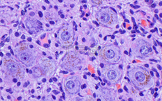
Light micrograph of ganglion cell bodies. Ganglion cells are part of the peripheral nervous system. Their cell bodies are characterized by large nucleoli within their nuclei (dark dots within paler circles) and abundant cytoplasm (paler blue substance around nuclei). Brown-yellow lipofuscin substance (small brown speckles) can also be seen in these ganglion cells. Haematoxylin and eosin stained tissue section. Magnification: 400x when printed at 10 centimetres.
| px | px | dpi | = | cm | x | cm | = | MB |
Details
Creative#:
TOP29978967
Source:
達志影像
Authorization Type:
RM
Release Information:
須由TPG 完整授權
Model Release:
N/A
Property Release:
N/A
Right to Privacy:
No
Same folder images:
pathologypathologicalmedicineanatomicalpathologypathologicalnervoussystemganglioncellsnerveganglionperipheralnervoussystemneuronneurologyneuropathologypathologicalbiologyhistologyhistologicalhistopathologypathologicallmlightmicroscopymagnifiedlightmicrographslidehematoxylinandeosinhaematoxylinandeosinhumanhumanbodymicroscopicanatomymicroanatomytissuecellcellscellbiologynormalbenignnobodyno-onemedicalmedicinehealthcare
anatomicalanatomyandandbenignbiologybiologybodycellcellcellscellseosineosinganglionganglionhaematoxylinhealthcarehematoxylinhistologicalhistologyhistopathologyhumanhumanlightlightlmmagnifiedmedicalmedicinemedicinemicroanatomymicrographmicroscopicmicroscopynervenervousnervousneurologyneuronneuropathologyno-onenobodynormalpathologicalpathologicalpathologicalpathologicalpathologypathologyperipheralslidesystemsystemtissue

 Loading