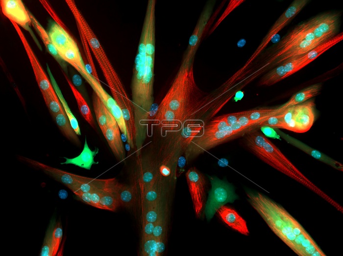
This image shows mouse muscle cells seen under a microscope. The cells have fused to form myotubes which have many nuclei (stained blue). The cells produced from mouse skeletal muscle stem cells with a harmless virus that made them glow green. The green color remained when the stem cells fused into myotubes. Some myotubes are stained red for a protein involved in muscle contraction (myosin heavy chain), a characteristic of mature muscle fibers. Researchers should use the same viral delivery system to genetically modify cells and assess how altering cell fusion impairs myotube growth. This image was the 2017 winner of the Federation of American Societies for Experimental Biology (FASEB) BioArt competition.
| px | px | dpi | = | cm | x | cm | = | MB |
Details
Creative#:
TOP28024647
Source:
達志影像
Authorization Type:
RM
Release Information:
須由TPG 完整授權
Model Release:
N/A
Property Release:
N/A
Right to Privacy:
No
Same folder images:

 Loading