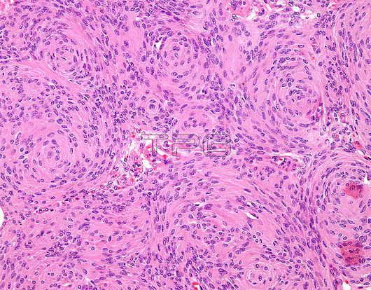
Meningothelial meningioma, light micrograph. Meningiomas are one of the commonest tumour types arising within the central nervous system and make up approximately 30% of primary intracranial tumours. 'Meningothelial' or 'syncytial' meningioma is one of the most common histologic subtypes of meningioma. It is a WHO grade I tumour. It is composed of spindly-looking cells with pink cytoplasm forming syncytial structures, whorls, and lobules separated by fibrovascular septa.
| px | px | dpi | = | cm | x | cm | = | MB |
Details
Creative#:
TOP25945383
Source:
達志影像
Authorization Type:
RM
Release Information:
須由TPG 完整授權
Model Release:
N/A
Property Release:
N/A
Right to Privacy:
No
Same folder images:
anaplasticanatomicpathologyarachnoidatypicalbraincancercancerouscarcinomacentralnervoussystemchromosome22cnsduraepilepsyhistologyhistopathologyhistopathologicalintracranialklf4lightmicroscopelightmicroscopylightmicrographleptomeningesmalignancymalignantmeningesmeningiomameningothelialneurofibromatosistype2nf2neurologicalneurologyneuropathologyoncologypathologypathologicalpsammomabodiespsammomatousrhabdoidseizurestraf7tumourtumourmedicinemedicalnobodyno-onehistologicallightmicrographlmtumor
222anaplasticanatomicarachnoidatypicalbodiesbraincancercancerouscarcinomacentralchromosomecnsduraepilepsyhistologicalhistologyhistopathologicalhistopathologyintracranialklf4leptomeningeslightlightlightlightlmmalignancymalignantmedicalmedicinemeningesmeningiomameningothelialmicrographmicrographmicroscopemicroscopynervousneurofibromatosisneurologicalneurologyneuropathologynf2no-onenobodyoncologypathologicalpathologypathologypsammomapsammomatousrhabdoidseizuressystemtraf7tumortumourtumourtype

 Loading