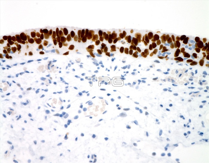
Urothelial carcinoma-in-situ (CIS), light micrograph stained for p53. Urothelial CIS is defined as the presence of overtly malignant cells with flat urothelium. The cancer's cells are discohesive and readily shed into the urine where they can be detected by cytological examination. Urothelial CIS is frequently multifocal and can show widespread involvement of the bladder lining with extension into the ureters and urethra. In the absence of treatment, about two-thirds to three-quarters of cases progress to invasive cancer. Microscopically, urothelial CIS shows full-thickness cytologic atypia consisting of enlarged hyperchromatic nuclei, prominent nucleoli and frequent mitotic figures. p53 is a useful immunostain to distinguish urothelial carcinoma-in-situ (diffusely positive) from reactive atypia (negative or weak non-specific staining).
| px | px | dpi | = | cm | x | cm | = | MB |
Details
Creative#:
TOP25499527
Source:
達志影像
Authorization Type:
RM
Release Information:
須由TPG 完整授權
Model Release:
N/A
Property Release:
N/A
Right to Privacy:
No
Same folder images:

 Loading