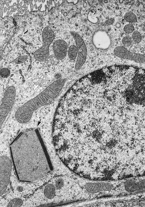
Electron micrograph of a proximal convoluted tubule cell from a rat. A peroxisome is visible near the cell base containing both crystals and a few circular profiles of cylinders in cross section. Despite their unusual appearance, these organelles can be identified as peroxisomes by positive staining for catalase and a negative reaction for acid phosphatase. Micrograph courtesy of Michael Barrett and Paul Heidger. THE CELL 2nd edition.
| px | px | dpi | = | cm | x | cm | = | MB |
Details
Creative#:
TOP22218883
Source:
達志影像
Authorization Type:
RM
Release Information:
須由TPG 完整授權
Model Release:
N/A
Property Release:
No
Right to Privacy:
No
Same folder images:

 Loading