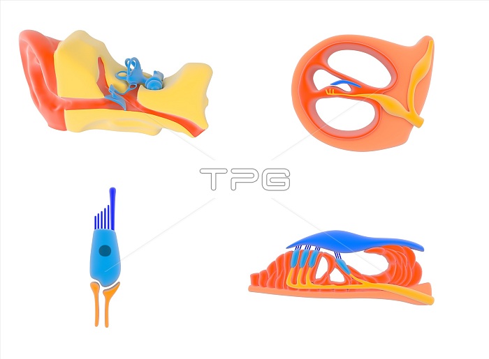
Ear and cochlear anatomy. Illustration of the human ear (upper left) and successively expanded views of the anatomy of the cochlea, the organ of hearing in the inner ear. The cochlea is one of the organs shown in blue at upper left. At upper right, a sectioned view through the cochlea shows the cochlear duct with the vestibular canal and typanic canal either side. At lower right, the organ of Corti consists of the tectorial membrane (blue), hair cells (dark blue and light blue), and sensory neurons (orange) leading to the auditory nerve. A hair cell is at lower left, with its cilia and sensory microvilli (dark blue). For this artwork with labels, see C023/8843.
| px | px | dpi | = | cm | x | cm | = | MB |
Details
Creative#:
TOP15984344
Source:
達志影像
Authorization Type:
RM
Release Information:
須由TPG 完整授權
Model Release:
N/A
Property Release:
N/A
Right to Privacy:
No
Same folder images:

 Loading