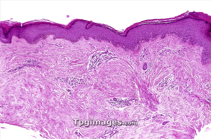
Keratoacanthoma. Light micrograph of a section through a keratoacanthoma nodule. The surface layers are across top, including the hard keratin layer. Below this there are patches of inflammation and white blood cells with stained nuclei (dark spots). The cause of a keratoacanthoma lesion is unknown but it has many features of a viral condition, and consists of a localised proliferation of squamous cells that forms a cratered nodule. The nodule grows over several weeks before gradually disappearing. However, the unsightly nodule is often surgically removed. The cause of keratoacanthoma is unknown, although exposure to sunlight appears to be a factor.
| px | px | dpi | = | cm | x | cm | = | MB |
Details
Creative#:
TOP06666495
Source:
達志影像
Authorization Type:
RM
Release Information:
須由TPG 完整授權
Model Release:
NO
Property Release:
NO
Right to Privacy:
No
Same folder images:

 Loading