filter
-
Brand
- By Category
- Direction
- Date Range
113Events
Pictures
Events

Editorial Electric Welding Institute of Academy of Sciences of Ukrainian SSR
- 2024-09-16
- 1

Editorial Artificial Blood Vessel
- 2024-08-22
- 2

Editorial EvX expo in Setia Alam, Malaysia - 21 Jul 2024
- 2024-07-22
- 3

Editorial NASA Set To Launch Next-Gen Solar Sail Spacecraft Into Deep Space Without Fuel Using ‘Stream Of Solar Wind’ From Sun
- 2024-04-18
- 5
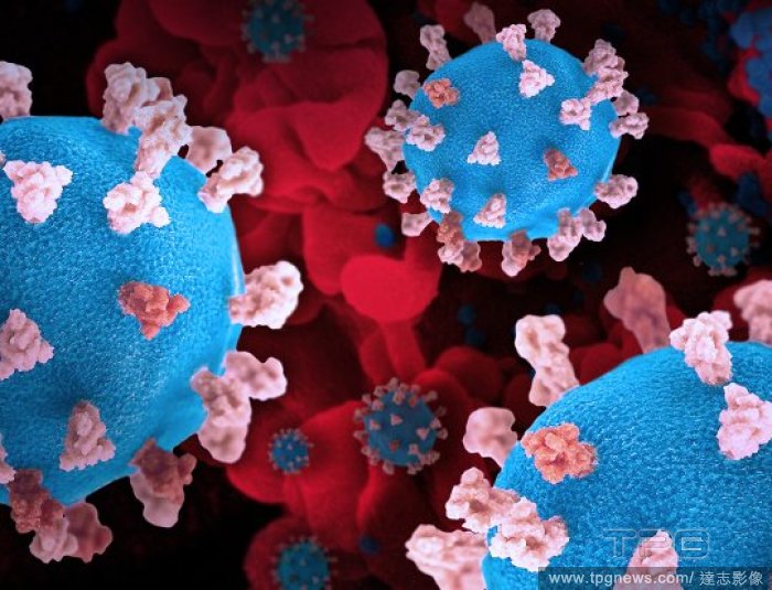
Editorial 52502267864_53cd1de5a9_o
- 2024-02-09
- 1

Editorial Photo illustration in Poland.
- 2024-01-31
- 1

Editorial Head of electron beam and laser welding
- 2023-12-28
- 1

Editorial Institute of Nuclear Physics, Siberian Branch of USSR Academy of Sciences
- 2023-12-06
- 1
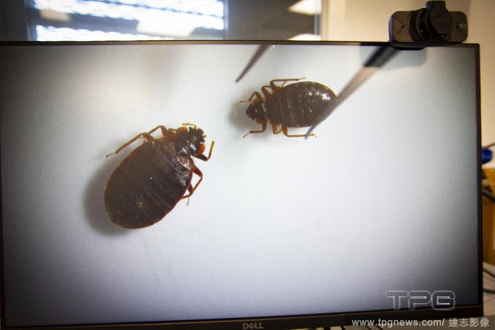
Editorial Bedbugs Study - Lyon, France - 06 Oct 2023
- 2023-10-10
- 8
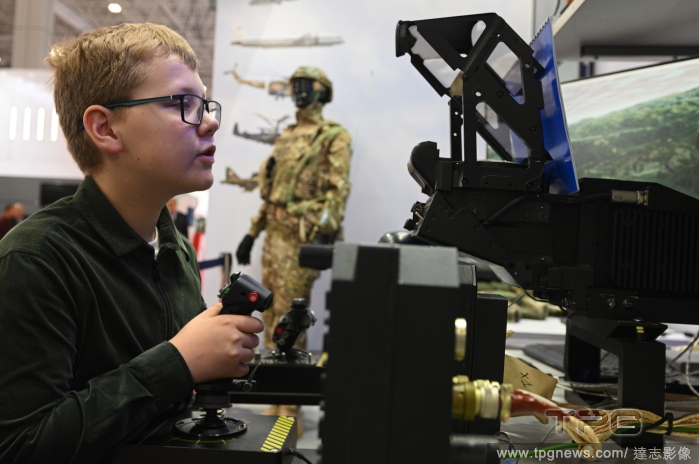
Editorial Russia Army Forum
- 2023-08-16
- 1
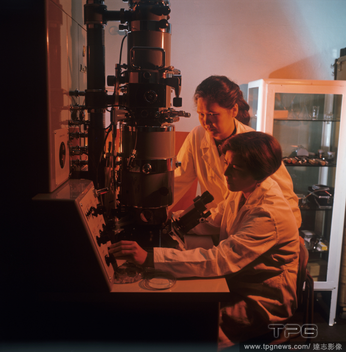
Editorial Satpayev Institute of Geological Sciences
- 2023-08-02
- 1

Editorial Satpayev Institute of Geological Sciences
- 2023-06-15
- 1

Editorial Scientists Solve Puzzle Of Red Streaks On Jupiter's Moon Europa
- 2023-02-21
- 3

Editorial Rocket Lab Launches Electron Rocket From USA
- 2023-01-28
- 6
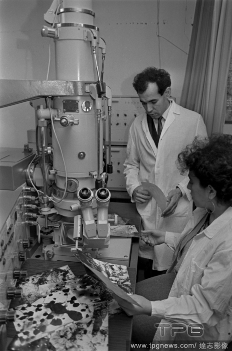
Editorial Special Design and Technological Bureau of Metal Science in Baku
- 2023-01-25
- 1
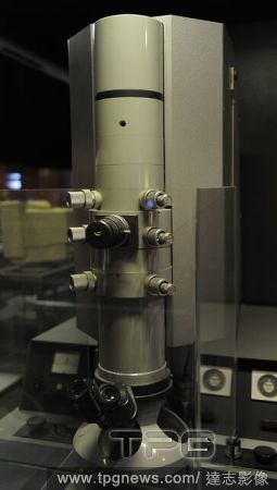
Editorial Transmission electron microscope "EM9". Signed: Carl Zeiss. 1964.
- 2022-12-24
- 1

Editorial Logos displayed on smartphones in Poland - 17 Dec 2022
- 2022-12-19
- 1

Editorial Joint Institute for Nuclear Research in Dubna
- 2022-12-14
- 1

Editorial Logos displayed on smartphones in Poland - 10 Dec 2022
- 2022-12-13
- 1
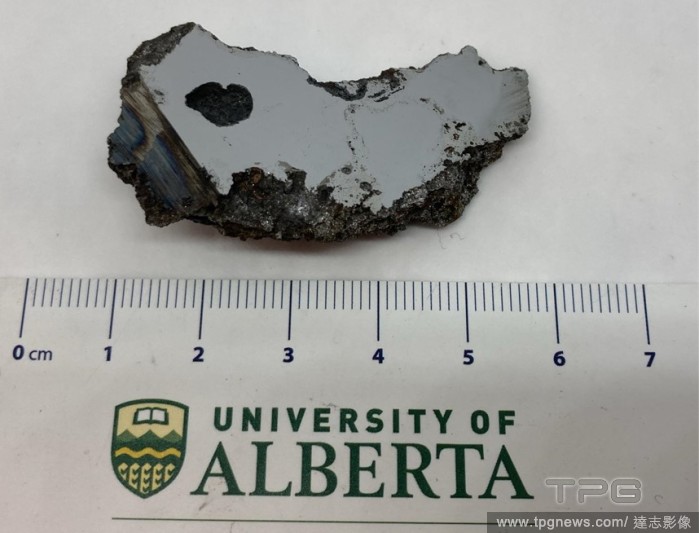
Editorial New Minerals Discovered In Meteorite Found In Somalia
- 2022-12-02
- 1
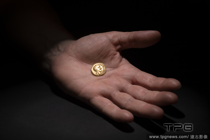
Editorial Coin Study Reveals Long-Lost Roman Emperor
- 2022-11-24
- 10

Editorial Elektron Color TVs
- 2022-11-19
- 1

Editorial Nano World Scientific Laboratory-Studio
- 2022-10-24
- 1
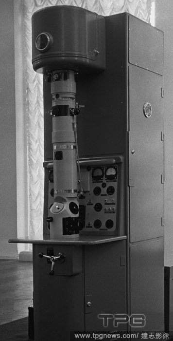
Editorial Unversal electron microscope UEM-100
- 2022-10-11
- 2
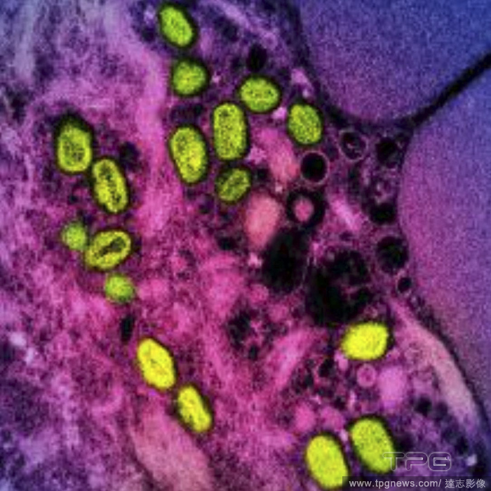
Editorial An undated image provided by the National Institute of Allergy and Infectious Diseases shows a colorized transmission electron micrograph of monkeypox particles (green) found within an infected cell (pink and purple), cultured in the laboratory. (National Institute of Allergy and Infectious Diseases via The New York Times)
- 2022-09-10
- 1
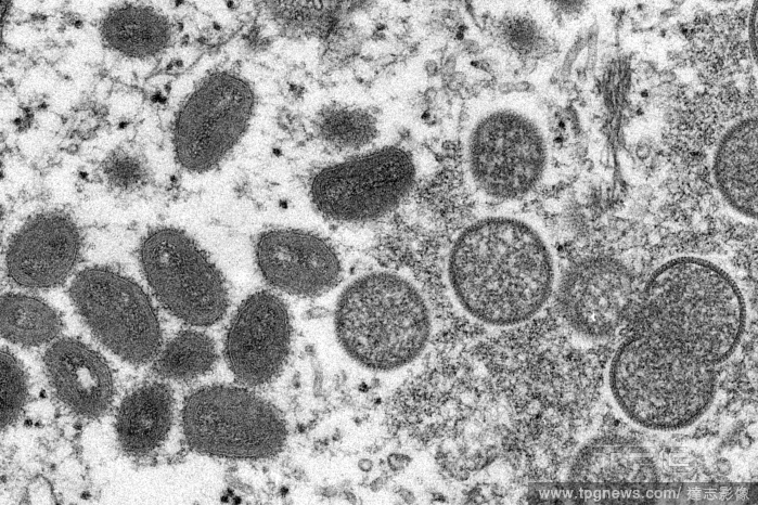
Editorial An electron microscope image provided by the U.S. Centers for Disease Control and Prevention shows oval-shaped, mature monkeybpox virions, left, and spherical, immature monkeypox virions, right, in 2003. (Centers for Disease Control and Prevention via The New York Times)
- 2022-06-17
- 1
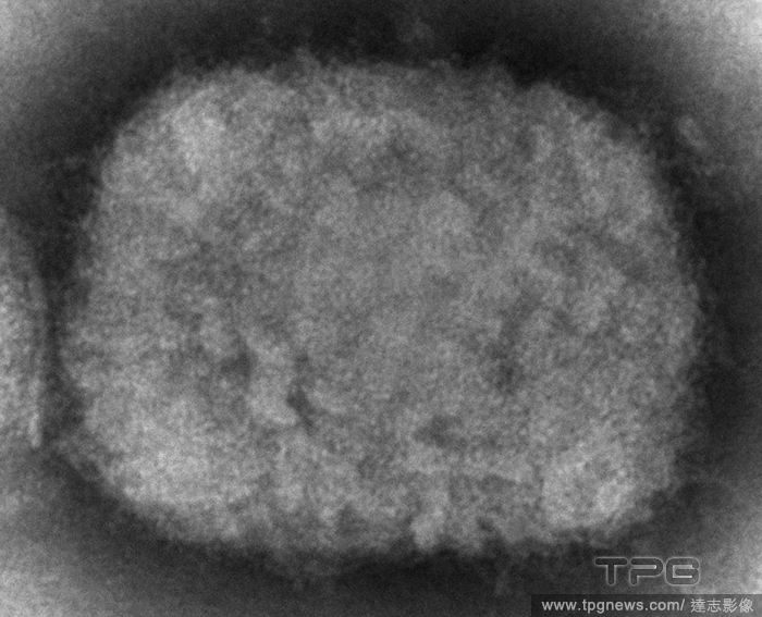
Editorial In an electron microscope image from the Centers for Disease Control and Prevention, a monkeypox virion in 2003 associated with a prairie dog outbreak that year. (Cynthia S. Goldsmith and Russell Regnery/Centers for Disease Control and Prevention via The New York Times)
- 2022-05-26
- 2
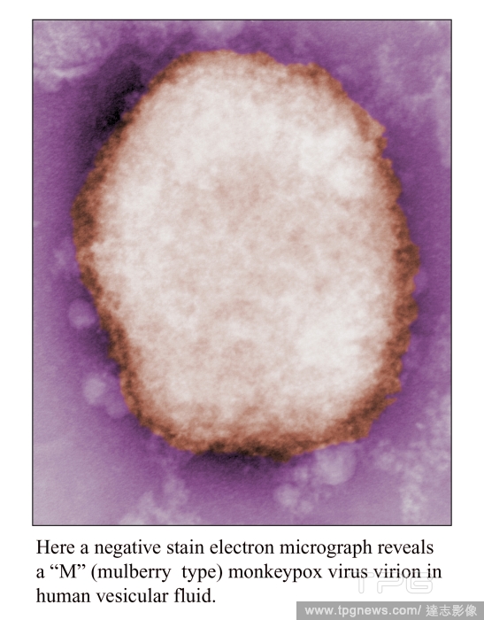
Editorial Monkeypox cases continue to rise around the world
- 2022-05-24
- 4
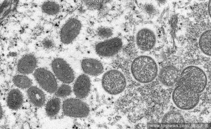
Editorial An undated electron microscopy image provided by the Centers for Disease Control and Prevention of mature monkeypox virions, left, and immature virions, right, from a sample of human skin associated with the 2003 outbreak of monkeypox in the U.S. (CDC via The New York Times)
- 2022-05-19
- 1
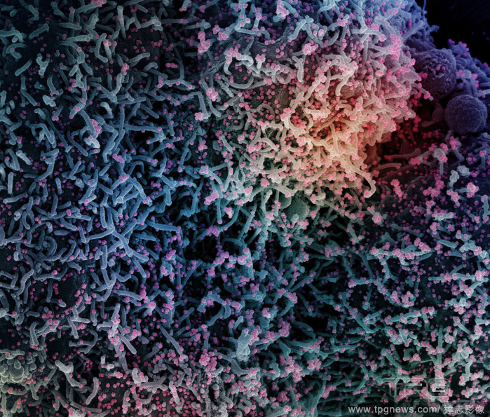
Editorial A colorized scanning electron micrograph of a cell infected with a coronavirus. (National Institutes of Health via The New York Times)
- 2022-02-01
- 1
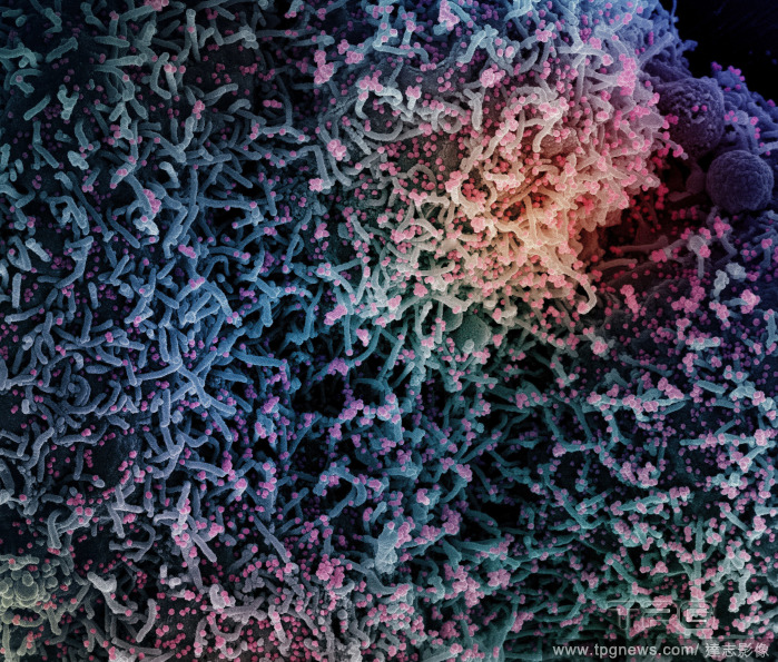
Editorial A colorized scanning electron micrograph of a cell infected with a coronavirus. (National Institutes of Health via The New York Times)
- 2022-01-25
- 1
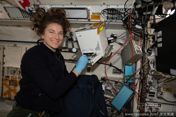
Editorial NASA Johnson
- 2022-01-22
- 1
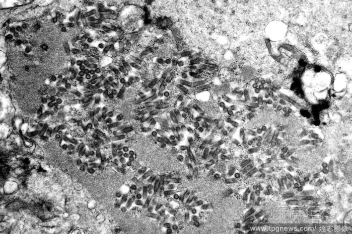
Editorial An electron microscope image provided by the Centers for Disease Control and Prevention shows rabies virions, dark and bullet-shaped, within an infected tissue sample. (F. A. Murphy/CDC via The New York Times)
- 2022-01-08
- 1

Editorial MA: Boston
- 2021-12-22
- 2

Editorial Press conference on research and preservation of Vincent van Gogh's painting Red Vineyard at Arles (Montmajour)
- 2021-12-20
- 22
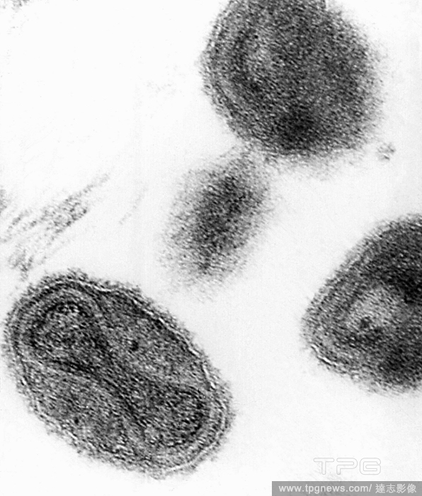
Editorial An electron micrograph of the smallpox virus taken in 1975. (Centers for Disease Control and Prevention via The New York Times)
- 2021-11-19
- 1
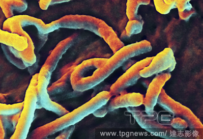
Editorial UPI Pictures of the Year 2014 -- NEWS AND FEATURES, Atlanta, Georgia, United States - 26 Nov 2014
- 2021-08-31
- 1

Editorial President Obama Presents the National Medals of Science & National Medals of Technology and Innovation in Washington, D.C, District of Columbia, United States - 20 Nov 2014
- 2021-08-31
- 2

Editorial Earrings, gold, forged, pressed, welded, granulated, electron (gold-silver alloy), total: height: 5.8 cm; diameter: 3.2 cm (arms); weight: 15 g, body jewelry, antique, The earring consists of a boat-shaped body with radially arranged conical arms. Of t...
- 2021-02-21
- 1
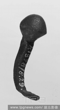
Editorial Quadrant bronze nail with double-electron, on top of round button. The tip is bent., Pen, metal, bronze, length: 3,5 cm, roman 1-300, Netherlands, North Brabant, Oss, Lith, Maas.
- 2021-02-20
- 1

Editorial applique, electron, silver, 2.9 x 3.4 cm, -500.
- 2021-02-20
- 1
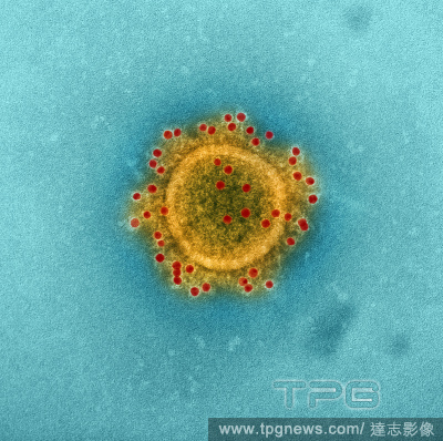
Editorial A transmission electron micrograph of the Middle East Respiratory Syndrome. (NIAID via The New York Times)
- 2021-02-16
- 1
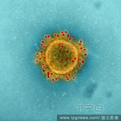
Editorial A transmission electron micrograph of the Middle East Respiratory Syndrome. (NIAID via The New York Times)
- 2021-02-10
- 1
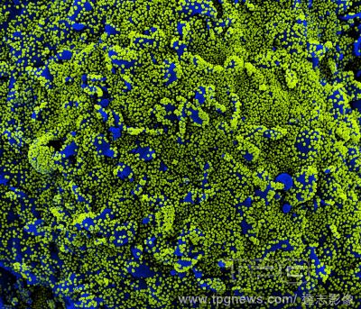
Editorial A colorized scanning electron micrograph of a cell (blue) heavily infected with coronavirus particles isolated from a patient sample in December 2020. (NIAID/National Institutes of Health via The New York Times)
- 2021-01-30
- 1
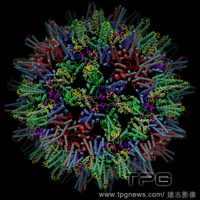
Editorial A graphic, made using cryo-electron microscopy, of a hepatitis B virus capsid where the protein is in red, green, yellow and blue (colors chosen to highlight capsid geometry) and a drug-like compound, HAP-TAMRA, in magenta. (Schlicksup, Wang, et al (2018) via The New York Times)
- 2021-01-26
- 1

Editorial A scanning electron micrograph of an apoptotic cell heavily infected with coronavirus particles. (National Institute of Allergy and Infectious Diseases via The New York Times)
- 2020-12-01
- 1

Editorial A scanning electron micrograph of an apoptotic cell heavily infected with coronavirus particles. (National Institute of Allergy and Infectious Diseases via The New York Times)
- 2020-11-28
- 1

Editorial Illustrations in Brazil - 18 Oct 2020
- 2020-10-19
- 2
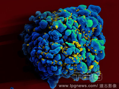
Editorial A colored scanning electron micrograph of a human T-cell, blue and green, infected with H.I.V., yellow. (NIAID via The New York Times)
- 2020-08-27
- 1

Editorial A coronavirus vaccine is on the horizon, thanks to a key discovery by these researchers
- 2020-08-14
- 1
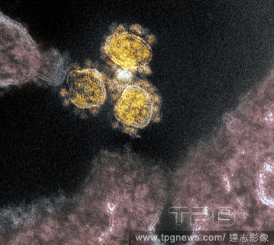
Editorial A colorized transmission electron microscope image of coronavirus. (NIAID via The New York Times)
- 2020-08-07
- 1
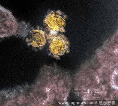
Editorial A colorized transmission electron microscope image of coronavirus. (NIAID via The New York Times)
- 2020-08-05
- 1
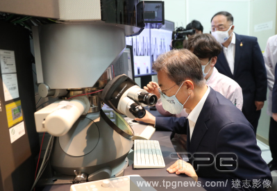
Editorial Moon visits SK hynix
- 2020-07-09
- 1
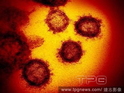
Editorial Truck driver posts regrets about party a day before dying of coronavirus
- 2020-07-04
- 1

Editorial In a photo from the National Institute of Allergy and Infectious Diseases, a colorized scanning electron micrograph of a dying cell infected with the coronavirus, with virus particles in red. (National Institute of Allergy and Infectious Diseases via The New York Times)
- 2020-07-03
- 1
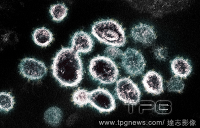
Editorial An electron microscope image provided by the National Institute of Allergy and Infectious Diseases shows SARS-CoV-2, the virus that causes COVID-19, isolated from a patient in the U.S. (NIAID via The New York Times)
- 2020-06-28
- 1
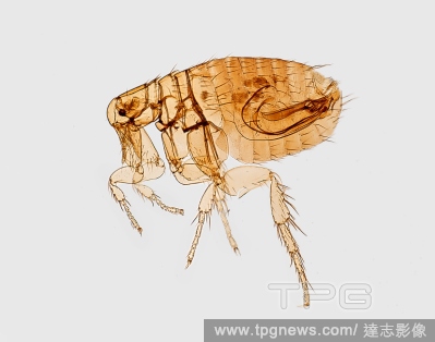
Editorial Flea Morphology
- 2020-06-09
- 3
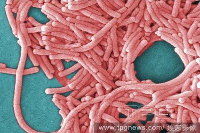
Editorial A digitally colorized scanning electron microscopic (SEM) image provided by Janice Haney Carr/Centers for Disease Control and Prevention shows Legionella pneumophila, the bacteria that causes Legionnaires’ Disease. (Janice Haney Carr/Centers for Disease Control and Prevention via The New York Times)
- 2020-05-21
- 1

Editorial Queen Maxima visit to the LUMC. Leiden, The Netherlands. 20 May 2020.
- 2020-05-21
- 26

Editorial Queen Maxima visit the LUMC
- 2020-05-21
- 27

Editorial A colored scanning electron micrograph provided by the National Institute of Allergy and Infectious Diseases shows a dying cell infected with the coronavirus, with viral particles in red. (National Institute of Allergy and Infectious Diseases via The New York Times)
- 2020-05-07
- 1
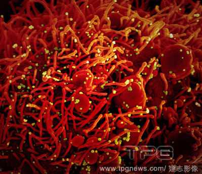
Editorial A scanning electron micrograph provided by the National Institute of Allergy and Infectious Diseases shows a dying cell infected with coronavirus particles. (National Institute of Allergy and Infectious Diseases via The New York Times)
- 2020-05-01
- 1
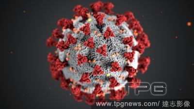
Editorial illustration of the Coronavirus (COVID-19), created at the Centers for Disease Control and Prevention (CDC)
- 2020-04-13
- 3
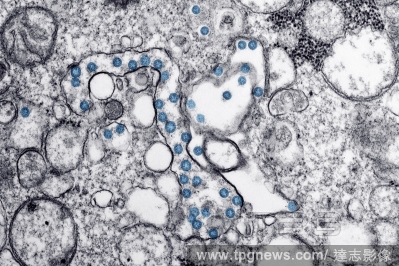
Editorial COVID-19 Coronavirus
- 2020-03-25
- 1
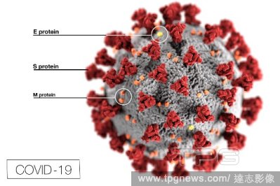
Editorial Microscopic Illustration of Coronavirus Disease 2019 (COVID-19)
- 2020-03-16
- 3
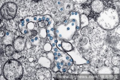
Editorial Microscopic View of Coronavirus Disease 2019 (COVID-19)
- 2020-03-16
- 1

Editorial Corona Virus in New York
- 2020-03-15
- 29

Editorial electron microscopic image of an isolate from the first U.S. case of COVID-19, formerly known as 2019-nCoV.
- 2020-03-14
- 2
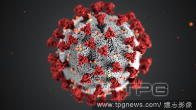
Editorial illustration of the Coronavirus (COVID-19), created at the Centers for Disease Control and Prevention (CDC)
- 2020-03-14
- 3
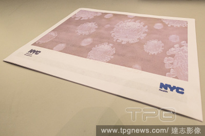
Editorial NY: NYC Mayor De Blasio and city officials hold table-top exercise on COVID-19 outbreak following NY's first confirmed case
- 2020-03-03
- 1

Editorial Microscopic view of COVID-19
- 2020-03-03
- 4
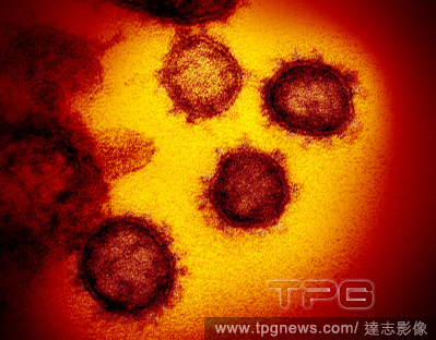
Editorial A transmission electron microscope image of 2019-nCoV, the virus that causes COVID-19, isolated from a patient in the U.S., emerging from the surface of cells cultured in a lab. (National Instittues of Health via The new York Times)
- 2020-03-01
- 1
 Loading