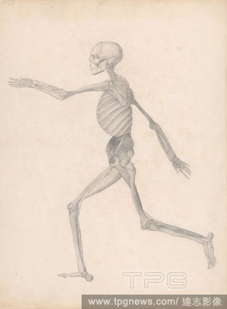
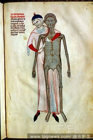
EditorialA Comparative Anatomical Exposition of the Structure of the Human Body with that of a Tiger and a Common Fowl: Human Figure, Lateral View (Finished study for an unpublished table, representing the last stage in the dissection). Date/Period: Between 179...
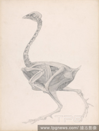
EditorialA Comparative Anatomical Exposition of the Structure of the Human Body with That of a Tiger and a Common Fowl: Fowl Body, Lateral View (Highly Finished Study for an Unpublished Table; Shows the Last Stage in Dissection). Date/Period: Between 1795 and 1...
EditorialA Comparative Anatomical Exposition of the Structure of the Human Body with That of a Tiger and a Common Fowl: Fowl Body, Lateral View (Highly Finished Study for an Unpublished Table; Shows the Last Stage in Dissection). Date/Period: Between 1795 and 1...
EditorialA Comparative Anatomical Exposition of the Structure of the Human Body with That of a Tiger and a Common Fowl: Fowl Body, Lateral View (Highly Finished Study for an Unpublished Table; Shows the Last Stage in Dissection). Date/Period: Between 1795 and 1...
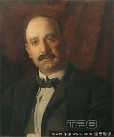
EditorialThomas Eakins, Portrait of A. Bryan Wall, 1904, oil on canvas, 24 1/4 in. x 20 3/16 in. (61.6 cm x 51.28 cm), At the outset of his career Thomas Eakins epitomized critical hopes for an art that would be both technically accomplished and distinctively n...
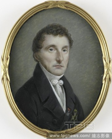
EditorialPieter de Riemer (1769-1831), professor of dissection and obstetrics, consultant surgeon of King William I, Portrait of Pieter de Riemer (1769-1831). Professor of dissection and obstetrics, consultant surgeon of King Willem I. Bust, to the right. Part ...
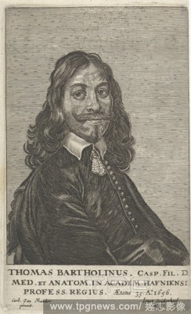
EditorialPortrait of Thomas Bartholinus, Danish physician and anatomist, at the age of 35. At the bottom in the margin a three-line text in Latin. Verso the title page for his book Anatomia: ofte, dissection of human body bodies, 1656, physician, doctor, Thomas...
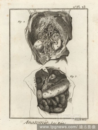
EditorialDissection of the human torso to reveal the kidneys. Copperplate engraving by Robert Benard after an illustration by Albrecht von Haller from Denis Diderot's Encyclopedia, Pellet, Geneva, 1779.
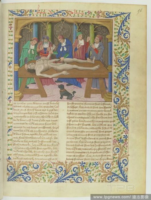
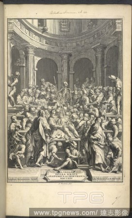
EditorialDissection. A. V. ... opera omnia anatomica et chirurgica cura. Lugduni Batavorum, 1725. Examination of a human corpse in front of an audience. Image taken from A. V. opera omnia anatomica et chirurgica cura H. Boerhaave et B. S. Albini. Originally p...
EditorialInterior of a pharmacy Title page for: J. Halmaal, Dissection of Amsterdam pharmacy, 1689, pharmacy, pharmaceutics, Jan Luyken (mentioned on object), Amsterdam, paper, etching, h 142 mm ? w 85 mm.
EditorialPieter de Riemer (1769-1831), professor of dissection and obstetrics, consultant surgeon of King William I, Portrait of Pieter de Riemer (1769-1831). Professor of dissection and obstetrics, consultant surgeon of King Willem I. Bust, to the right. Part ...
EditorialPortrait of Thomas Bartholinus, Danish physician and anatomist, at the age of 35. At the bottom in the margin a three-line text in Latin. Verso the title page for his book Anatomia: ofte, dissection of human body bodies, 1656, physician, doctor, Thomas...
EditorialJean-Baptiste Andr? Gautier-Dagoty (French, 1740 - 1786). Dissection of a Lizard, Plate 15, 1756. Mezzotint printed in colors on laid paper. Plate: 212 mm x 146 mm (8.35 in. x 5.75 in.).
EditorialA Comparative Anatomical Exposition of the Structure of the Human Body with That of a Tiger and a Common Fowl: Fowl Body, Lateral View (Highly Finished Study for an Unpublished Table; Shows the Last Stage in Dissection). Date/Period: Between 1795 and 1...
EditorialAn anatomical dissection. Le Otto Tavole Anatomiche con cinquanta figure in. Roma, 1750. An anatomical dissection being performed. Image taken from Le Otto Tavole Anatomiche con cinquanta figure in foglio delineate per compimento dell:opera del B. Eus...
EditorialDissection. A. V. ... opera omnia anatomica et chirurgica cura. Lugduni Batavorum, 1725. Examination of a human corpse in front of an audience. Image taken from A. V. opera omnia anatomica et chirurgica cura H. Boerhaave et B. S. Albini. Originally p...
EditorialA Comparative Anatomical Exposition of the Structure of the Human Body with That of a Tiger and a Common Fowl: Fowl Body, Lateral View (Highly Finished Study for an Unpublished Table; Shows the Last Stage in Dissection). Date/Period: Between 1795 and 1...
EditorialDissection of the human torso to reveal the kidneys. Copperplate engraving by Robert Benard after an illustration by Albrecht von Haller from Denis Diderot's Encyclopedia, Pellet, Geneva, 1779.
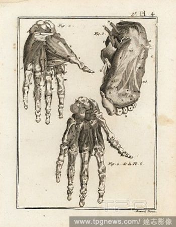
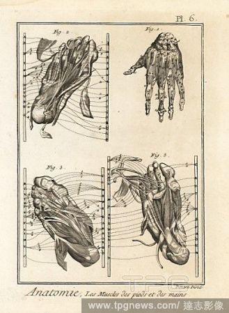
EditorialDissection of the human hand and foot to reveal muscle, ligament and bone. Copperplate engraving by Robert Benard from Denis Diderot's Encyclopedia, Pellet, Geneva, 1779.
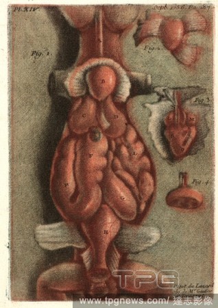
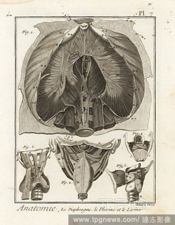
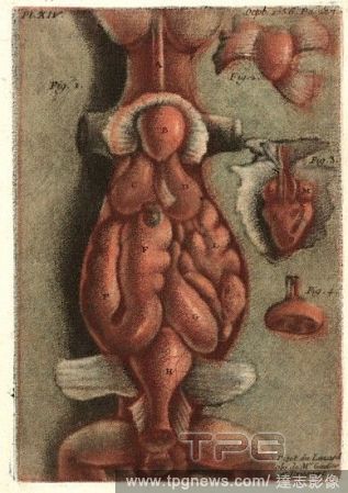
EditorialDissection of the human diaphragm, chest cavity, and details of the pharynx and larynx. Copperplate engraving by Robert Benard after an illustration by Albrecht von Haller from Denis Diderot's Encyclopedia, Pellet, Geneva, 1779.
EditorialDissection of the muscles in the hand and foot. Copperplate engraving by Robert Benard from Denis Diderot's Encyclopedia, Pellet, Geneva, 1779.
EditorialJean-Baptiste Andr? Gautier-Dagoty (French, 1740 - 1786). Dissection of a Lizard, Plate 15, 1756. Mezzotint printed in colors on laid paper. Plate: 212 mm x 146 mm (8.35 in. x 5.75 in.).


 Loading