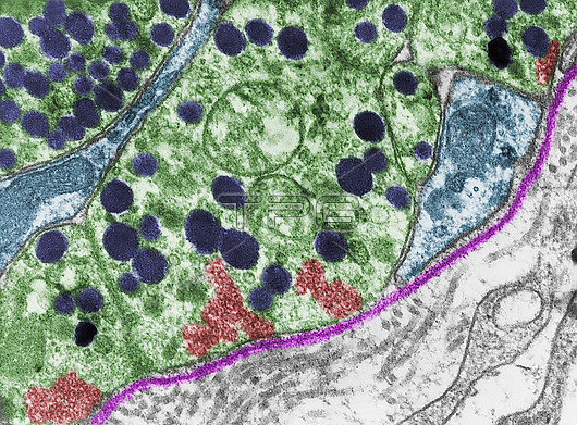
Coloured transmission electron micrograph (TEM) showing terminal axons (green) of hypothalamic neurosecretory neurons (nerve cells) with secretory granules (dark blue) and clusters of clear synaptic vesicles joined to a plasma membrane. A basement membrane (pink) and the thin processes of pituicytes (light blue) are also seen.
| px | px | dpi | = | cm | x | cm | = | MB |
Details
Creative#:
TOP30082563
Source:
達志影像
Authorization Type:
RM
Release Information:
須由TPG 完整授權
Model Release:
N/A
Property Release:
N/A
Right to Privacy:
No
Same folder images:
pituitaryneurohypophysisendocrineglandendocrinologyglandglandularherringposteriorpituitaryhistologicalhistologyhormonehumanbodyhypophysisparsnervosapituitarypituitaryglandneurosecretoryhistologyhistologicalbiologybiologicaltemtransmissionelectronmicrographmicroscopynobodyno-onehealthynormalcolouredcoloredfalse-colouredfalse-colored
biologicalbiologybodycoloredcolouredelectronendocrineendocrinologyfalse-coloredfalse-colouredglandglandglandglandularhealthyherringhistologicalhistologicalhistologyhistologyhormonehumanhypophysismicrographmicroscopynervosaneurohypophysisneurosecretoryno-onenobodynormalparspituitarypituitarypituitarypituitaryposteriortemtransmission

 Loading