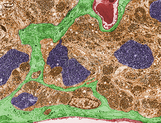
Coloured transmission electron micrograph (TEM) of the posterior pituitary gland (neurohypophysis). The neurohypophysis parenchyma (brown) shows many axons full of neurosecretory granules. There are several large expansions of these axons, known as Herring bodies (dark blue). The connective tissue stroma, with blood capillaries (red), is green.
| px | px | dpi | = | cm | x | cm | = | MB |
Details
Creative#:
TOP30082561
Source:
達志影像
Authorization Type:
RM
Release Information:
須由TPG 完整授權
Model Release:
N/A
Property Release:
N/A
Right to Privacy:
No
Same folder images:
pituitaryneurohypophysisendocrineglandendocrinologyglandglandularherringposteriorpituitaryhistologicalhistologyhormonehumanbodyhypophysisparsnervosapituitarypituitaryglandneurosecretoryoxytocinvasopressinhistologyhistologicalbiologybiologicaltemtransmissionelectronmicrographmicroscopynobodyno-onehealthynormalcolouredcoloredfalse-colouredfalse-colored
biologicalbiologybodycoloredcolouredelectronendocrineendocrinologyfalse-coloredfalse-colouredglandglandglandglandularhealthyherringhistologicalhistologicalhistologyhistologyhormonehumanhypophysismicrographmicroscopynervosaneurohypophysisneurosecretoryno-onenobodynormaloxytocinparspituitarypituitarypituitarypituitaryposteriortemtransmissionvasopressin

 Loading