
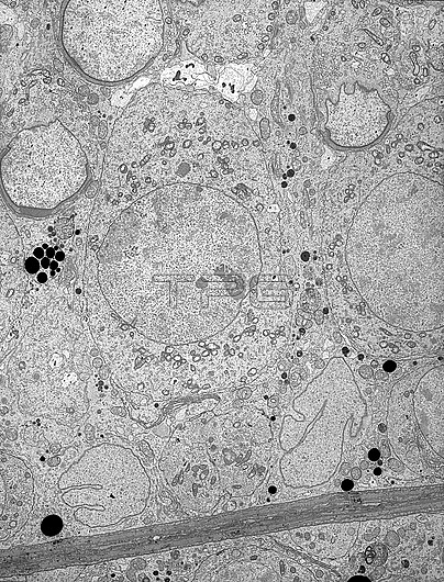
Transmission electron micrograph (TEM) of the ultrastructure of the seminiferous epithelium showing two Sertoli cell nuclei adjacent to the basal lamina. At centre is a pachytene primary spermatocyte above which are three spermatids in the early phase of elongation. The spermatids have developed an acrosome which covers the basally-facing aspect of their nuclei. Magnification: x2,000 when height printed at 10cm.
| px | px | dpi | = | cm | x | cm | = | MB |
Details
Creative#:
TOP27779074
Source:
達志影像
Authorization Type:
RM
Release Information:
須由TPG 完整授權
Model Release:
N/A
Property Release:
N/A
Right to Privacy:
No
Same folder images:
TestisseminiferousepitheliumspermatogenesisSertolicellspermatocyteprimaryspermatocytemalereproductionmalereproductivesystemgermcellmalegermcellacrosomeultrastructureelectronmicrographelectronmicroscopytransmissionelectronmicrographcellultrastructurecellbiologytemnobodyno-oneblackandwhitemonochromebiologicalcytologycytological
SertoliTestisacrosomeandbiologicalbiologyblackcellcellcellcellcellcytologicalcytologyelectronelectronelectronepitheliumgermgermmalemalemalemicrographmicrographmicroscopymonochromeno-onenobodyprimaryreproductionreproductiveseminiferousspermatocytespermatocytespermatogenesissystemtemtransmissionultrastructureultrastructurewhite
 Loading