
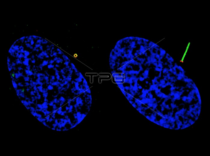
Cilia-based defects cancer research. Structured illumination microscopy (SIM) imaging of two human retinal pigment epithelium (RPE) cells, showing ciliogenesis progression (the formation of cilia). Defects in ciliary formation and function are linked to several human diseases, including cancer. Ciliogenesis starts from docking pre-ciliary vesicles to the distal appendage of the mother centriole. Those docked vesicles fuse to form a larger ciliary vesicle and then extend to the ciliary membrane. At left, GFP-Smoothened (green) vesicles are docked at the mother centriole/basal body distal appendage (marked by CEP164 in red). At right, ciliary GFP-Smoothened (green) is localized on the ciliary membrane. The cell nuclei are shown in blue.
| px | px | dpi | = | cm | x | cm | = | MB |
Details
Creative#:
TOP19460885
Source:
達志影像
Authorization Type:
RM
Release Information:
須由TPG 完整授權
Model Release:
N/A
Property Release:
N/A
Right to Privacy:
No
Same folder images:
 Loading