
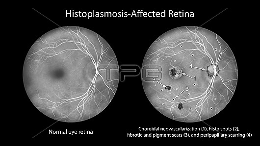
Illustration of a retina affected by presumed ocular histoplasmosis syndrome as seen in fluorescein angiography. The retina shows choroidal neovascularization, histo spots, fibrotic and pigment scars, peripapillary scarring.
| px | px | dpi | = | cm | x | cm | = | MB |
Details
Creative#:
TPG34965984
Source:
達志影像
Authorization Type:
RF
Release Information:
須由TPG 完整授權
Model Release:
N/A
Property Release:
N/A
Right to Privacy:
No
Same folder images:
histoplasmosishistoplasmaretinaabnormalabnormalitiesanomaliesconditiondisorderdiagnosticfundoscopyfundusopticdiscretinalretinalanatomyretinoscopyophthalmoscopyophthalmoscopeillustrationmedicaleyeeyeexaminationophthalmologyophthalmologicalpathologyophthalmicanatomydiscoculardiseasefungalchronicfungiblackbackgroundhanddrawndrawingsyndromecapsulatumhistospotsscarpigmentedspotschoroidalneovascularizationscarringfibroticfibrosisfluoresceinangiographyangiogramscarringblackandwhitemonochrometext
abnormalabnormalitiesanatomyanatomyandangiogramangiographyanomaliesbackgroundblackblackcapsulatumchoroidalchronicconditiondiagnosticdiscdiscdiseasedisorderdrawingdrawnexaminationeyeeyefibrosisfibroticfluoresceinfundoscopyfundusfungalfungihandhistohistoplasmahistoplasmosisillustrationmedicalmonochromeneovascularizationocularophthalmicophthalmologicalophthalmologyophthalmoscopeophthalmoscopyopticpathologypigmentedretinaretinalretinalretinoscopyscarscarringscarringspotsspotssyndrometextwhite
 Loading