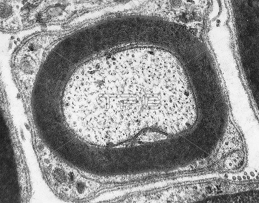
Transmission electron micrograph (TEM) of a cross-sectioned peripheral myelinated nerve fibre. At centre is the axon with many cross-sectioned neurofilaments. Around the axon, the laminated structure of myelin appears as alternating dark lines (protein layers) separated by unstained zones (the lipid hydrocarbon chains). Outside the myelin, the Schwann cell cytoplasm (showing the mesaxon) surrounded by a basement membrane, can be seen.
| px | px | dpi | = | cm | x | cm | = | MB |
Details
Creative#:
TOP30082577
Source:
達志影像
Authorization Type:
RM
Release Information:
須由TPG 完整授權
Model Release:
N/A
Property Release:
N/A
Right to Privacy:
No
Same folder images:

 Loading