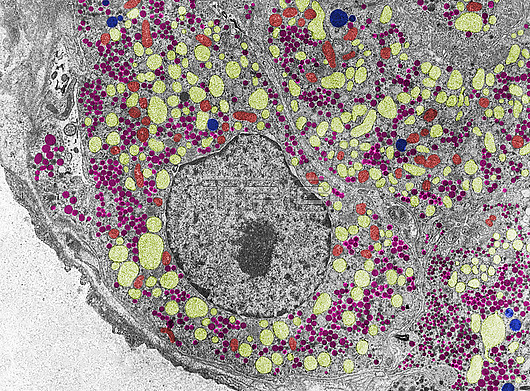
Pituitary gland. Coloured transmission electron micrograph (TEM) of a gonadotropic cell of a castrated animal showing dark follicle stimulating hormone (FSH) and luteinising hormone (LH) granules (pink) and large swellings of the rough endoplasmic reticulum (RER, light yellow) due to the castration. Mitochondria are red and lysosomes blue. The lack of sex hormones due to castration causes hypertrophy of gonadotroph cells that secrete increased amounts of FSH and LH to counteract the lack of sex hormones.
| px | px | dpi | = | cm | x | cm | = | MB |
Details
Creative#:
TOP30082549
Source:
達志影像
Authorization Type:
RM
Release Information:
須由TPG 完整授權
Model Release:
N/A
Property Release:
N/A
Right to Privacy:
No
Same folder images:
adenohypophysisanteriorpituitarycastrationcellcellcellbiologycytologyelectronelectronmicroscopyendoplasmicreticulumfinestructurefsh-lhgranulesgonadotropiccellhistologyhypophysislysosomesmicroscopyorganellespituitarypituitaryglandrerrerswellingstemultrastructurehistologyhistologicalbiologybiologicaltemtransmissionelectronmicrographmicroscopynobodyno-onehealthynormalcolouredcoloredfalse-colouredfalse-colored
adenohypophysisanteriorbiologicalbiologybiologycastrationcellcellcellcellcoloredcolouredcytologyelectronelectronelectronendoplasmicfalse-coloredfalse-colouredfinefsh-lhglandgonadotropicgranuleshealthyhistologicalhistologyhistologyhypophysislysosomesmicrographmicroscopymicroscopymicroscopyno-onenobodynormalorganellespituitarypituitarypituitaryrerrerreticulumstructureswellingstemtemtransmissionultrastructure

 Loading