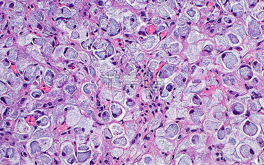
Light micrograph of signet ring cell carcinoma. Signet ring cell carcinoma is formed of cells with cytoplasmic mucin (pale blue-grey) and nuclei (dark blue dots) which are pushed to the periphery of the cell. Signet ring cell carcinoma most commonly occurs in the gastrointestinal tract. Haematoxylin and eosin stained tissue section. Magnification: 200x when printed at 10 centimetres.
| px | px | dpi | = | cm | x | cm | = | MB |
Details
Creative#:
TOP30048856
Source:
達志影像
Authorization Type:
RM
Release Information:
須由TPG 完整授權
Model Release:
N/A
Property Release:
N/A
Right to Privacy:
No
Same folder images:
pathologypathologicalmedicineanatomicalpathologypathologicalsignetringcellscancercancerouscarcinomagastrointestinalcanceroncologyoncologicaltumortumourmasssignetringcellcarcinomasurgicalpathologypathologicalmucinvacuolebiologybiologicalhistologyhistologicalhistopathologypathologicallmlightmicroscopymagnifiedlightmicrographslidehematoxylinandeosinhaematoxylinandeosinhumanhumanbodymicroscopicanatomymicroanatomytissuecellcellscellbiologynobodyno-onemedicalmedicinehealthcare
anatomicalanatomyandandbiologicalbiologybiologybodycancercancercancerouscarcinomacarcinomacellcellcellcellscellseosineosingastrointestinalhaematoxylinhealthcarehematoxylinhistologicalhistologyhistopathologyhumanhumanlightlightlmmagnifiedmassmedicalmedicinemedicinemicroanatomymicrographmicroscopicmicroscopymucinno-onenobodyoncologicaloncologypathologicalpathologicalpathologicalpathologicalpathologypathologypathologyringringsignetsignetslidesurgicaltissuetumortumourvacuole

 Loading