
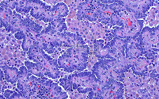
Light micrograph of lung adenocarcinoma. The lung cancer cells (dark blue) form glandular structures and are admixed with inflammatory cells, mostly histiocytes (lighter pink-purple). Haematoxylin and eosin stained tissue section. Magnification: 200x when printed at 10 centimetres.
| px | px | dpi | = | cm | x | cm | = | MB |
Details
Creative#:
TOP29978987
Source:
達志影像
Authorization Type:
RM
Release Information:
須由TPG 完整授權
Model Release:
N/A
Property Release:
N/A
Right to Privacy:
No
Same folder images:
pathologypathologicalmedicineanatomicalpathologypathologicallunglungcanceradenocarcinomatumortumourmassoncologyoncologicalpulmonologypulmonarypathologypathologicalthoracicpathologypathologicalcarcinomabiologyhistologyhistologicalhistopathologypathologicallmlightmicroscopymagnifiedlightmicrographslidehematoxylinandeosinhaematoxylinandeosinhumanhumanbodymicroscopicanatomymicroanatomytissuecellcellscellbiologynobodyno-onemedicalmedicinehealthcare
adenocarcinomaanatomicalanatomyandandbiologybiologybodycancercarcinomacellcellcellseosineosinhaematoxylinhealthcarehematoxylinhistologicalhistologyhistopathologyhumanhumanlightlightlmlunglungmagnifiedmassmedicalmedicinemedicinemicroanatomymicrographmicroscopicmicroscopyno-onenobodyoncologicaloncologypathologicalpathologicalpathologicalpathologicalpathologicalpathologypathologypathologypathologypulmonarypulmonologyslidethoracictissuetumortumour
 Loading