
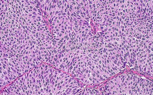
Light micrograph of high grade papillary urothelial carcinoma. The tumour cells are composed of oval-round nuclei (purple) and have eosinophilic cytoplasm (pink). The cells form papillae, the cores of which are seen as the dark pink-red strands between the tumour cells. Haematoxylin and eosin stained tissue section. Magnification: 200x when printed at 10 centimetres.
| px | px | dpi | = | cm | x | cm | = | MB |
Details
Creative#:
TOP29978974
Source:
達志影像
Authorization Type:
RM
Release Information:
須由TPG 完整授權
Model Release:
N/A
Property Release:
N/A
Right to Privacy:
No
Same folder images:
pathologypathologicalmedicineanatomicalpathologypathologicalbladdercancerurothelialcarcinomapapillaryurothelialcarcinomacancercancerousoncologyoncologicaltumortumourmassgenitourinarypathologypathologicalurologyurologicalbiologyhistologyhistologicalhistopathologypathologicallmlightmicroscopymagnifiedlightmicrographslidehematoxylinandeosinhaematoxylinandeosinhumanhumanbodymicroscopicanatomymicroanatomytissuecellcellscellbiologydiseasenobodyno-onemedicalmedicinehealthcare
anatomicalanatomyandandbiologybiologybladderbodycancercancercancerouscarcinomacarcinomacellcellcellsdiseaseeosineosingenitourinaryhaematoxylinhealthcarehematoxylinhistologicalhistologyhistopathologyhumanhumanlightlightlmmagnifiedmassmedicalmedicinemedicinemicroanatomymicrographmicroscopicmicroscopyno-onenobodyoncologicaloncologypapillarypathologicalpathologicalpathologicalpathologicalpathologypathologypathologyslidetissuetumortumoururologicalurologyurothelialurothelial
 Loading