
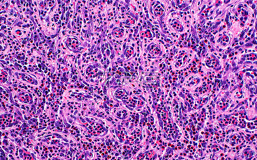
Light micrograph of granulation tissue. Granulation tissue is made up of a mixture of inflammatory cells (most of the dark blue-dots) and small blood vessels (pink circular structures) within a fibrous (pink) to myxoid (pale blue) stroma. Haematoxylin and eosin stained tissue section. Magnification: 200x when printed at 10 centimetres.
| px | px | dpi | = | cm | x | cm | = | MB |
Details
Creative#:
TOP29978970
Source:
達志影像
Authorization Type:
RM
Release Information:
須由TPG 完整授權
Model Release:
N/A
Property Release:
N/A
Right to Privacy:
No
Same folder images:
pathologypathologicalmedicineanatomicalpathologypathologicalgranulationtissueinflammationbiologyhistologyhistologicalhistopathologypathologicallmlightmicroscopymagnifiedlightmicrographslidehematoxylinandeosinhaematoxylinandeosinhumanhumanbodymicroscopicanatomymicroanatomytissuecellcellscellbiologynobodyno-onemedicalmedicinehealthcare
anatomicalanatomyandandbiologybiologybodycellcellcellseosineosingranulationhaematoxylinhealthcarehematoxylinhistologicalhistologyhistopathologyhumanhumaninflammationlightlightlmmagnifiedmedicalmedicinemedicinemicroanatomymicrographmicroscopicmicroscopyno-onenobodypathologicalpathologicalpathologicalpathologypathologyslidetissuetissue
 Loading