
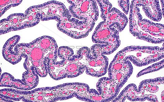
Light micrograph of fallopian tube fimbria. The fimbria are finger-like projections at the ends of the fallopian tubes which function to help move an ovulated ovum down the fallopian tube lumen. The fimbria seen in this image are lined by a layer of epithelial cells (blue-purple) and contain stroma (white and pink) and small blood vessels (darker red circles). Haematoxylin and eosin stained tissue section. Magnification: 100x when printed at 10 centimetres.
| px | px | dpi | = | cm | x | cm | = | MB |
Details
Creative#:
TOP29978958
Source:
達志影像
Authorization Type:
RM
Release Information:
須由TPG 完整授權
Model Release:
N/A
Property Release:
N/A
Right to Privacy:
No
Same folder images:
pathologypathologicalmedicineanatomicalpathologypathologicalfallopiantubefimbriareproductionfemalereproductivesystemreproductiongynecologygynecologicalgynaecologygynaecologicalgynecologicpathologypathologicaladnexabiologyhistologyhistologicalhistopathologypathologicallmlightmicroscopymagnifiedlightmicrographslidehematoxylinandeosinhaematoxylinandeosinhumanhumanbodymicroscopicanatomymicroanatomytissuecellcellscellbiologynormalbenignnobodyno-onemedicalmedicinehealthcare
adnexaanatomicalanatomyandandbenignbiologybiologybodycellcellcellseosineosinfallopianfemalefimbriagynaecologicalgynaecologygynecologicgynecologicalgynecologyhaematoxylinhealthcarehematoxylinhistologicalhistologyhistopathologyhumanhumanlightlightlmmagnifiedmedicalmedicinemedicinemicroanatomymicrographmicroscopicmicroscopyno-onenobodynormalpathologicalpathologicalpathologicalpathologicalpathologypathologypathologyreproductionreproductionreproductiveslidesystemtissuetube
 Loading