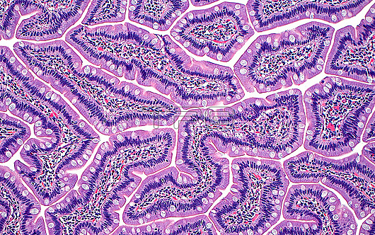
Light micrograph of a section of duodenal villi. The duodenal villous cells include goblet cells (white dots) and columnar epithelial cell nuclei (thin blue lines parallel to each other). Duodenal villi can produce interesting patterns, as seen in this image, when cut at different cross sections and angles. Haematoxylin and eosin stained tissue section. Magnification: 100x when printed at 10 centimetres.
| px | px | dpi | = | cm | x | cm | = | MB |
Details
Creative#:
TOP29978955
Source:
達志影像
Authorization Type:
RM
Release Information:
須由TPG 完整授權
Model Release:
N/A
Property Release:
N/A
Right to Privacy:
No
Same folder images:
pathologypathologicalmedicineanatomicalpathologypathologicalgastrointestinalgiduodenumvillidigestivesystembowelsmallboweldigestiondigestivetractgastrointestinaltractintestinegobletcellssurgicalpathologypathologicalbiologyhistologyhistologicalhistopathologypathologicallmlightmicroscopymagnifiedlightmicrographslidehematoxylinandeosinhaematoxylinandeosinhumanhumanbodymicroscopicanatomymicroanatomytissuecellcellscellbiologynormalbenignnobodyno-onemedicalmedicinehealthcare
anatomicalanatomyandandbenignbiologybiologybodybowelbowelcellcellcellscellsdigestiondigestivedigestiveduodenumeosineosingastrointestinalgastrointestinalgigoblethaematoxylinhealthcarehematoxylinhistologicalhistologyhistopathologyhumanhumanintestinelightlightlmmagnifiedmedicalmedicinemedicinemicroanatomymicrographmicroscopicmicroscopyno-onenobodynormalpathologicalpathologicalpathologicalpathologicalpathologypathologypathologyslidesmallsurgicalsystemtissuetracttractvilli

 Loading