
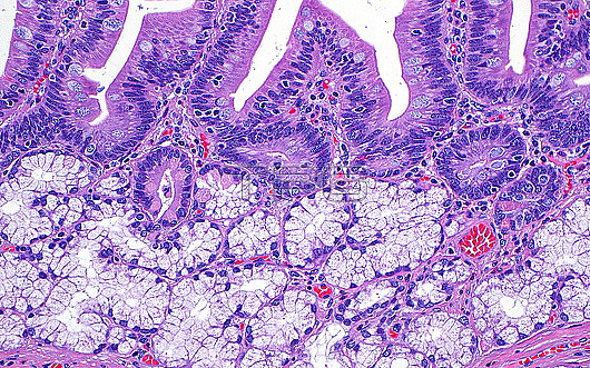
Light micrograph of Brunner?™s glands. Brunner?™s glands (light grey-white structures at bottom half of picture) are present in the duodenum and underlie the duodenal villi (top half of picture) Haematoxylin and eosin stained tissue section. Magnification: 200x when printed at 10 centimetres.
| px | px | dpi | = | cm | x | cm | = | MB |
Details
Creative#:
TOP29978953
Source:
達志影像
Authorization Type:
RM
Release Information:
須由TPG 完整授權
Model Release:
N/A
Property Release:
N/A
Right to Privacy:
No
Same folder images:
pathologypathologicalmedicineanatomicalpathologypathologicalduodenumbrunnerglandsglandgastrointestinalgidigestivesystemgastrointestinalpathologypathologicalsurgicalpathologypathologicalbiologyhistologyhistologicalhistopathologypathologicallmlightmicroscopymagnifiedlightmicrographslidehematoxylinandeosinhaematoxylinandeosinhumanhumanbodymicroscopicanatomymicroanatomytissuecellcellscellbiologynormalbenignnobodyno-onemedicalmedicinehealthcare
anatomicalanatomyandandbenignbiologybiologybodybrunnercellcellcellsdigestiveduodenumeosineosingastrointestinalgastrointestinalgiglandglandshaematoxylinhealthcarehematoxylinhistologicalhistologyhistopathologyhumanhumanlightlightlmmagnifiedmedicalmedicinemedicinemicroanatomymicrographmicroscopicmicroscopyno-onenobodynormalpathologicalpathologicalpathologicalpathologicalpathologicalpathologypathologypathologypathologyslidesurgicalsystemtissue
 Loading