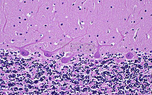
Light micrograph of cerebellum Purkinje neurons. The distinctive Purkinje neuron cell bodies (central image) are located between the granular cell layer (darker blue, bottom half of picture), and the molecular layer (top light pink half of picture). Dendrites (thin projections) branch out from the cell bodies into the molecular layer. Haematoxylin and eosin stained tissue section. Magnification: 200x when printed at 10 centimetres.
| px | px | dpi | = | cm | x | cm | = | MB |
Details
Creative#:
TOP29978926
Source:
達志影像
Authorization Type:
RM
Release Information:
須由TPG 完整授權
Model Release:
N/A
Property Release:
N/A
Right to Privacy:
No
Same folder images:
pathologypathologicalmedicineanatomicalpathologypathologicalcerebellumbrainpurkinjeneuronscerebellumlayerscerebellarcortexcentralnervoussystemnervoussystemneurologyneuropathologypathologicalneuroanatomynervecellsneuronsbiologyhistologyhistologicalhistopathologypathologicallmlightmicroscopymagnifiedlightmicrographslidehematoxylinandeosinstainhumanhumanbodymicroscopicanatomymicroanatomytissuecellcellscellbiologynormalbenignnobodyno-onemedicalmedicinehealthcare
anatomicalanatomyandbenignbiologybiologybodybraincellcellcellscellscentralcerebellarcerebellumcerebellumcortexeosinhealthcarehematoxylinhistologicalhistologyhistopathologyhumanhumanlayerslightlightlmmagnifiedmedicalmedicinemedicinemicroanatomymicrographmicroscopicmicroscopynervenervousnervousneuroanatomyneurologyneuronsneuronsneuropathologyno-onenobodynormalpathologicalpathologicalpathologicalpathologicalpathologypathologypurkinjeslidestainsystemsystemtissue

 Loading