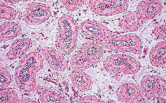
Light micrograph of atrophic seminiferous tubules in a testis. The seminiferous tubules show thickened basement membranes (dark pink) and absent or only immature germ cells (blue dots). The tubules are present within an oedematous stroma (light blue-white background). A few small blood vessels are also seen in the stroma. Haematoxylin and eosin stained tissue section. Magnification: 100x when printed at 10 centimetres.
| px | px | dpi | = | cm | x | cm | = | MB |
Details
Creative#:
TOP29978910
Source:
達志影像
Authorization Type:
RM
Release Information:
須由TPG 完整授權
Model Release:
N/A
Property Release:
N/A
Right to Privacy:
No
Same folder images:
pathologypathologicalmedicineanatomicalpathologypathologicaltestismalemalereproductivesystemreproductionatrophyseminiferoustubulesgenitourinarysystemurologyurologicalgenitourinarypathologypathologicalbiologyhistologyhistologicalhistopathologypathologicallmlightmicroscopymagnifiedlightmicrographslidehematoxylinandeosinhaematoxylinandeosinhumanhumanbodymicroscopicanatomymicroanatomytissuecellcellscellbiologyfertilityinfertilitynobodyno-onemedicalmedicinehealthcare
anatomicalanatomyandandatrophybiologybiologybodycellcellcellseosineosinfertilitygenitourinarygenitourinaryhaematoxylinhealthcarehematoxylinhistologicalhistologyhistopathologyhumanhumaninfertilitylightlightlmmagnifiedmalemalemedicalmedicinemedicinemicroanatomymicrographmicroscopicmicroscopyno-onenobodypathologicalpathologicalpathologicalpathologicalpathologypathologypathologyreproductionreproductiveseminiferousslidesystemsystemtestistissuetubulesurologicalurology

 Loading