
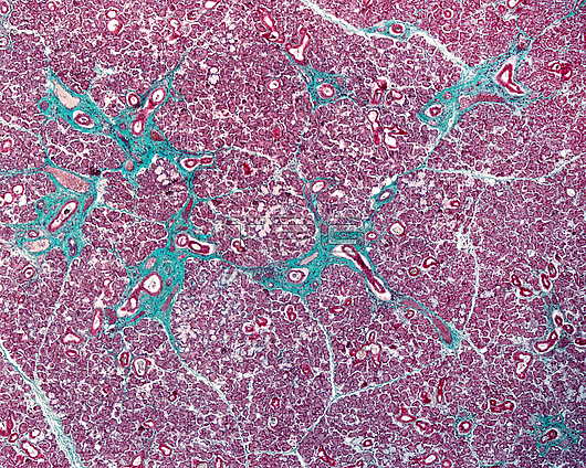
Light micrograph of a human submandibular gland stained with the trichrome method. The parenchyma shows very abundant serous tubule-acini and fewer mixed tubule-acini. The connective tissue septa are highlighted by the trichrome method, which stains the collagen fibres green.
| px | px | dpi | = | cm | x | cm | = | MB |
Details
Creative#:
TOP29874386
Source:
達志影像
Authorization Type:
RM
Release Information:
須由TPG 完整授權
Model Release:
N/A
Property Release:
N/A
Right to Privacy:
No
Same folder images:
 Loading