
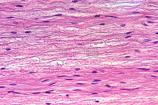
Light micrograph of a longitudinal section of a nerve showing numerous parallel myelinated nerve fibres. Most of the nuclei observed are of Schwann cells. In three fibres, the nodes of Ranvier can be seen.
| px | px | dpi | = | cm | x | cm | = | MB |
Details
Creative#:
TOP29874377
Source:
達志影像
Authorization Type:
RM
Release Information:
須由TPG 完整授權
Model Release:
N/A
Property Release:
N/A
Right to Privacy:
No
Same folder images:
nervefiberfibreranviernodeofranvierhistologylightmicroscopemicrographmicroscopymyelinmyelinatednervefiberfibrenervenervoussystemnervoustissueneuroanatomyneurohistologyneuroimagingneurologyneurosciencepnsschwanncelltissuenobodyno-onelightmicrographlmmicroscopynormalhealthyhistologyhistologicalanatomyanatomicalbiologybiological
anatomicalanatomybiologicalbiologycellfiberfiberfibrefibrehealthyhistologicalhistologyhistologylightlightlmmicrographmicrographmicroscopemicroscopymicroscopymyelinmyelinatednervenervenervenervousnervousneuroanatomyneurohistologyneuroimagingneurologyneuroscienceno-onenobodynodenormalofpnsranvierranvierschwannsystemtissuetissue
 Loading