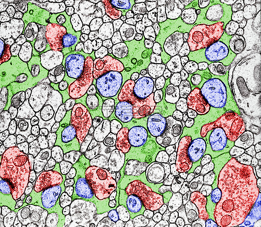
Coloured transmission electron micrograph (TEM) of the cerebellar molecular layer showing axodendritic synapses between granule cell axons (red) and dendritic spines of Purkinje cells (blue). Bergmann cell glia processes (blue) are surrounding and isolating the synapses.
| px | px | dpi | = | cm | x | cm | = | MB |
Details
Creative#:
TOP29845906
Source:
達志影像
Authorization Type:
RM
Release Information:
須由TPG 完整授權
Model Release:
N/A
Property Release:
N/A
Right to Privacy:
No
Same folder images:
synapsemolecularlayercerebellarcortexaxodendriticaxongranulecellspinepurkinjedendritebiologycellcytologymicroscopybergmannsynaptictemultrastructurevesiclemitochondriasmoothendoplasmicreticulumbiologybiologicalhistologyhistologicalnobodyno-onetransmissionelectronmicrographtemmicroscopyhealthynormalcolouredcoloredfalse-colouredfalse-colored
axodendriticaxonbergmannbiologicalbiologybiologycellcellcerebellarcoloredcolouredcortexcytologydendriteelectronendoplasmicfalse-coloredfalse-colouredgranulehealthyhistologicalhistologylayermicrographmicroscopymicroscopymitochondriamolecularno-onenobodynormalpurkinjereticulumsmoothspinesynapsesynaptictemtemtransmissionultrastructurevesicle

 Loading