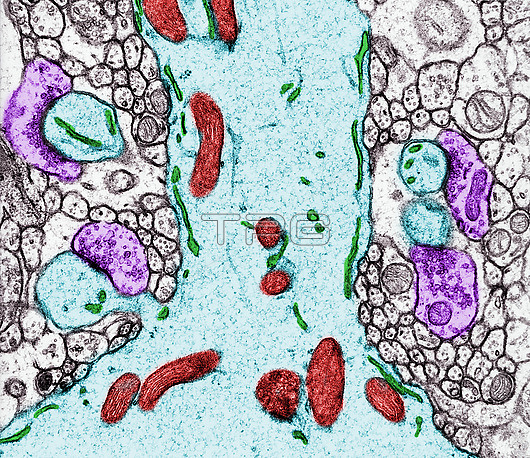
Coloured transmission electron micrograph (TEM) of a Purkinje cell dendrite showing some organelles; mitochondria (red) and smooth endoplasmic reticulum (green). At bottom left, a dendritic spine can be seen receiving a synapse from a granule cell axon (purple). Another three dendritic spine heads (the section plane does not pass through their necks) also show synapses.
| px | px | dpi | = | cm | x | cm | = | MB |
Details
Creative#:
TOP29845904
Source:
達志影像
Authorization Type:
RM
Release Information:
須由TPG 完整授權
Model Release:
N/A
Property Release:
N/A
Right to Privacy:
No
Same folder images:
synapsemolecularlayercerebellarcortexaxodendriticaxongranulecellspinepurkinjedendritebiologycellcytologymicroscopybergmannsynaptictemultrastructurevesiclemitochondriasmoothendoplasmicreticulumbiologybiologicalhistologyhistologicalnobodyno-onetransmissionelectronmicrographtemmicroscopyhealthynormalcolouredcoloredfalse-colouredfalse-colored
axodendriticaxonbergmannbiologicalbiologybiologycellcellcerebellarcoloredcolouredcortexcytologydendriteelectronendoplasmicfalse-coloredfalse-colouredgranulehealthyhistologicalhistologylayermicrographmicroscopymicroscopymitochondriamolecularno-onenobodynormalpurkinjereticulumsmoothspinesynapsesynaptictemtemtransmissionultrastructurevesicle

 Loading