
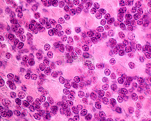
Light micrograph of the granular layer of the cerebellar cortex stained with haematoxylin and eosin. Under the Purkinje cells (at top) is the granular layer, which has many nuclei of granule cells, one of the smallest but most abundant brain neurons (nerve cells). Among the granule cells nuclei, the cerebellar glomeruli appear as pink areas without nuclei.
| px | px | dpi | = | cm | x | cm | = | MB |
Details
Creative#:
TOP29820565
Source:
達志影像
Authorization Type:
RM
Release Information:
須由TPG 完整授權
Model Release:
N/A
Property Release:
N/A
Right to Privacy:
No
Same folder images:
cerebellarcortexgreymattergraymattercerebellumbrainpurkinjecortexfibrefibergranularlayercnscentralnervoussystemhistologicalhistologylightmicrographlightmicroscopemicroscopymolecularlayerwhitematternervousneurohistologyneurologicalneurologylightmicrographlightmicroscopemicroscopyhistologyhistologicallmnobodyno-onenormalhealthybiologybiological
biologicalbiologybraincentralcerebellarcerebellumcnscortexcortexfiberfibregranulargraygreyhealthyhistologicalhistologicalhistologyhistologylayerlayerlightlightlightlightlmmattermattermattermicrographmicrographmicroscopemicroscopemicroscopymicroscopymolecularnervousnervousneurohistologyneurologicalneurologyno-onenobodynormalpurkinjesystemwhite
 Loading