
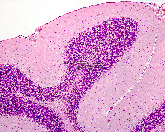
Light micrograph of a cerebellar folium showing the layers of cerebellar cortex: an external molecular layer, a Purkinje cell layer, a granular layer and the central axis of white matter.
| px | px | dpi | = | cm | x | cm | = | MB |
Details
Creative#:
TOP29820556
Source:
達志影像
Authorization Type:
RM
Release Information:
須由TPG 完整授權
Model Release:
N/A
Property Release:
N/A
Right to Privacy:
No
Same folder images:
cerebellarcortexgreymattergraymattercerebellumbrainpurkinjecortexfibrefibergranularlayercnscentralnervoussystemhistologicalhistologylightmicrographlightmicroscopemicroscopymolecularlayerwhitematternervousneurohistologyneurologicalneurologylightmicrographlightmicroscopemicroscopyhistologyhistologicallmnobodyno-onenormalhealthybiologybiological
biologicalbiologybraincentralcerebellarcerebellumcnscortexcortexfiberfibregranulargraygreyhealthyhistologicalhistologicalhistologyhistologylayerlayerlightlightlightlightlmmattermattermattermicrographmicrographmicroscopemicroscopemicroscopymicroscopymolecularnervousnervousneurohistologyneurologicalneurologyno-onenobodynormalpurkinjesystemwhite
 Loading