
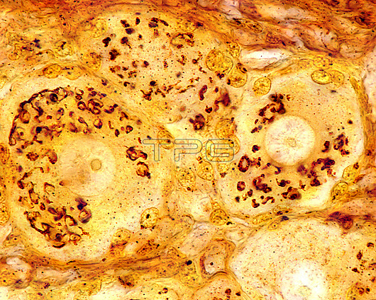
Light micrograph of pseudounipolar neurons(nerve cells) of a dorsal root ganglion stained with Cajals formol-uranium silver method to highlight the Golgi apparatus. It appears as a brown network located in the neuron cell body around the nucleus, except in the axon hillock, which appears as a clear zone devoid of Golgi apparatus components.
| px | px | dpi | = | cm | x | cm | = | MB |
Details
Creative#:
TOP29814545
Source:
達志影像
Authorization Type:
RM
Release Information:
須由TPG 完整授權
Model Release:
N/A
Property Release:
N/A
Right to Privacy:
No
Same folder images:
golgiapparatusgolgisilveraxonhillockbiologycellcellbodycajaldictyosomedorsalrootganglionhistologylightmicroscopelightmicrographhealthynormalbiologybiologicalhistologicalnervecellneuronnobodyno-onemicroscopynervoussystemnervoustissueneuroanatomyneurohistologyneuroimagingneurologyneuronneurosciencepseudounipolarformol-uraniumsilver
apparatusaxonbiologicalbiologybiologybodycajalcellcellcelldictyosomedorsalformol-uraniumgangliongolgigolgihealthyhillockhistologicalhistologylightlightmicrographmicroscopemicroscopynervenervousnervousneuroanatomyneurohistologyneuroimagingneurologyneuronneuronneuroscienceno-onenobodynormalpseudounipolarrootsilversilversystemtissue
 Loading