
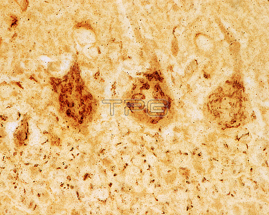
Cerebellar cortex. Light micrograph of Purkinje cells stained with Cajals formol-uranium silver method to show the large Golgi apparatus of these neurons (nerve cells). It appears as a brown network located in the soma of the Purkinje cells. The granular layer shows a stippled appearance due to staining of the small Golgi apparatus in the cerebellar granule cells.
| px | px | dpi | = | cm | x | cm | = | MB |
Details
Creative#:
TOP29814533
Source:
達志影像
Authorization Type:
RM
Release Information:
須由TPG 完整授權
Model Release:
N/A
Property Release:
N/A
Right to Privacy:
No
Same folder images:
cerebellumbraingolgiapparatuspurkinjecellscentralnervoussystemcerebellarcortexcnsdictyosomegolgihistologylightmicroscopelightmicrographhealthynormalbiologybiologicalhistologicalnervecellneuronnobodyno-onemicroscopynervoussystemnervoustissueneurohistologyneuronneurosciencepurkinjetissueformol-uraniumsilver
apparatusbiologicalbiologybraincellcellscentralcerebellarcerebellumcnscortexdictyosomeformol-uraniumgolgigolgihealthyhistologicalhistologylightlightmicrographmicroscopemicroscopynervenervousnervousnervousneurohistologyneuronneuronneuroscienceno-onenobodynormalpurkinjepurkinjesilversystemsystemtissuetissue
 Loading