
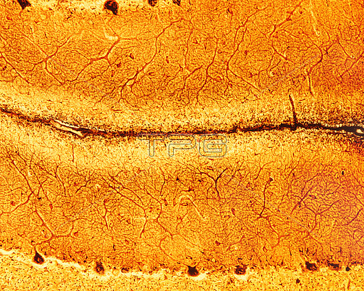
Light micrograph of immature cerebellar cortex impregnated with a silver method that demonstrates the Golgi apparatus in the Purkinje cells and also the dendritic tree of these neurons (nerve cells). The clear band on the top of the molecular layer is the external granular layer (characteristic of immature cerebellum) where the cerebellar granular neuron progenitors are located.
| px | px | dpi | = | cm | x | cm | = | MB |
Details
Creative#:
TOP29814529
Source:
達志影像
Authorization Type:
RM
Release Information:
須由TPG 完整授權
Model Release:
N/A
Property Release:
N/A
Right to Privacy:
No
Same folder images:
cerebellumbraindevelopmentexternalgranularlayergolgiapparatuspurkinjecellscentralnervoussystemcerebellarcortexcnsdictyosomegolgihistologylightmicroscopelightmicrographhealthynormalbiologybiologicalhistologicalnervecellneuronnobodyno-onemicroscopynervoussystemnervoustissueneurohistologyneuronneurosciencepurkinjetissueformol-uraniumsilver
apparatusbiologicalbiologybraincellcellscentralcerebellarcerebellumcnscortexdevelopmentdictyosomeexternalformol-uraniumgolgigolgigranularhealthyhistologicalhistologylayerlightlightmicrographmicroscopemicroscopynervenervousnervousnervousneurohistologyneuronneuronneuroscienceno-onenobodynormalpurkinjepurkinjesilversystemsystemtissuetissue
 Loading