
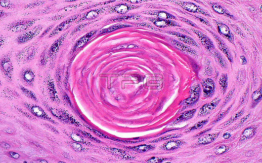
Light micrograph of keratin at the surface of skin. This image shows keratin material (hot pink) at the surface of skin surrounded by squamous cells. The skin epidermal (surface lining layer) cells produce keratin that forms the outermost protective layer of normal skin. Haematoxylin and eosin stained tissue section. Magnification: 400x when printed at 10cm.
| px | px | dpi | = | cm | x | cm | = | MB |
Details
Creative#:
TOP29676321
Source:
達志影像
Authorization Type:
RM
Release Information:
須由TPG 完整授權
Model Release:
N/A
Property Release:
N/A
Right to Privacy:
No
Same folder images:
pathologypathologicalmedicinemedicaldiseasedisorderconditionabnormalunhealthyanatomicpathologyskinkeratinskinlayersepidermisskinepidermisdermatologydermatopathologyhistopathologicalhistopathologymicroscopylmlightmicrographslidehematoxylinandeosinhaematoxylinandeosinhumanbodyanatomytissuecellsnormalhealthynobodyno-one
abnormalanatomicanatomyandandbodycellsconditiondermatologydermatopathologydiseasedisordereosineosinepidermisepidermishaematoxylinhealthyhematoxylinhistopathologicalhistopathologyhumankeratinlayerslightlmmedicalmedicinemicrographmicroscopyno-onenobodynormalpathologicalpathologypathologyskinskinskinslidetissueunhealthy
 Loading