
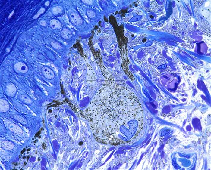
Chromatophore, light micrograph. A chromatophore of the skin of the melanophore class located in the dermis below the superficial epidermis seen at left. Thousands of pigment granules of melanin are in the cell body and also dispersed into the cytoplasmic extensions that terminate at the epidermal-dermal junction. An irregular shaped nucleus is in the cell body. These cells are very large, the cell body 50-70 micrometres and the extensions up to 100 micrometres. Melanophores are found in the skin of animals that use colour for camouflage (reptiles, fish, amphibians, cephalopods) and absorb light giving brown/black colouration. Surrounding the cell and scattered in the dermis are layers of plaques of iridophore cells with tiny blue-green refractile crystalloids that give blue colourations to the skin. Epoxy resin section. Toluidine blue stain. Magnification: x900 when printed at 10cm.
| px | px | dpi | = | cm | x | cm | = | MB |
Details
Creative#:
TOP29389370
Source:
達志影像
Authorization Type:
RM
Release Information:
須由TPG 完整授權
Model Release:
Not Available
Property Release:
Not Available
Right to Privacy:
No
Same folder images:
 Loading