
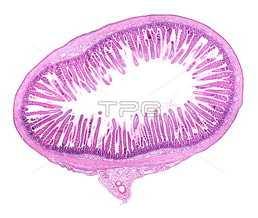
Light micrograph of a cross-sectioned rat small intestine. The mucosa layer shows abundant villi, finger-like projections that extend into the lumen. Outside the mucosa, the submucosa and the muscular layer can be seen. The mesentery, with blood vessels (round), is at bottom centre.
| px | px | dpi | = | cm | x | cm | = | MB |
Details
Creative#:
TOP29223587
Source:
達志影像
Authorization Type:
RM
Release Information:
須由TPG 完整授權
Model Release:
Not Available
Property Release:
Not Available
Right to Privacy:
No
Same folder images:
 Loading