
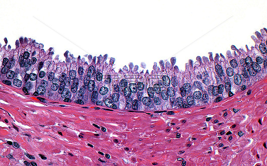
Light micrograph of prostate intraepithelial neoplasia (PIN) in a prostate gland. The presence of PIN indicates a heightened risk for prostate cancer. PIN cells have hyperchromatic (dark blue) nuclei with a pseudostratified (layer-like) arrangement. The PIN cells also show blebs into the luminal space of the prostate gland (small rounded projections into the white space). The PIN cells do not invade (as would true cancer cells) and are contained within the prostate gland which itself lies within connective stromal tissue (pink-red area in bottom half of image). Haematoxylin and eosin stained tissue section. Magnification: x400 when printed at 10cm wide.
| px | px | dpi | = | cm | x | cm | = | MB |
Details
Creative#:
TOP29066948
Source:
達志影像
Authorization Type:
RM
Release Information:
須由TPG 完整授權
Model Release:
Not Available
Property Release:
Not Available
Right to Privacy:
No
Same folder images:
 Loading