
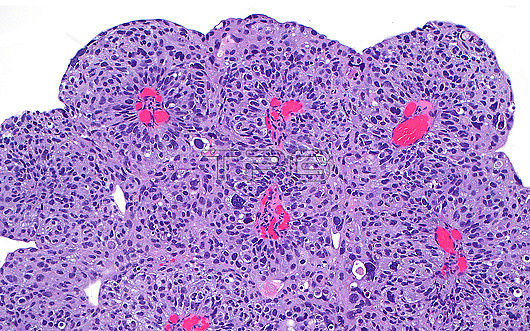
Light micrograph of papillary urothelial carcinoma (bladder cancer). The cancer cells are arranged in complex papillary structures, which are structures that have cores containing blood vessels (bright red). Haematoxylin and eosin stained tissue section. Magnification: x100 when printed at 10cm wide.
| px | px | dpi | = | cm | x | cm | = | MB |
Details
Creative#:
TOP29066947
Source:
達志影像
Authorization Type:
RM
Release Information:
須由TPG 完整授權
Model Release:
Not Available
Property Release:
Not Available
Right to Privacy:
No
Same folder images:
pathologymedicinepathologicalmedicalanatomicpathologyurinarysystembladdercancerurinarybladderurothelialcarcinomapapillaryhighgradegenitourinarypathologyurologytransurethralresectionbiologyhistologyhistologicalhistopathologymicroscopylmlightmicrographslideHematoxylinHaematoxylinandeosinstainnobodyno-onehumanbodyanatomytissuecellswhitebackground
HaematoxylinHematoxylinanatomicanatomyandbackgroundbiologybladderbladderbodycancercarcinomacellseosingenitourinarygradehighhistologicalhistologyhistopathologyhumanlightlmmedicalmedicinemicrographmicroscopyno-onenobodypapillarypathologicalpathologypathologypathologyresectionslidestainsystemtissuetransurethralurinaryurinaryurologyurothelialwhite
 Loading