
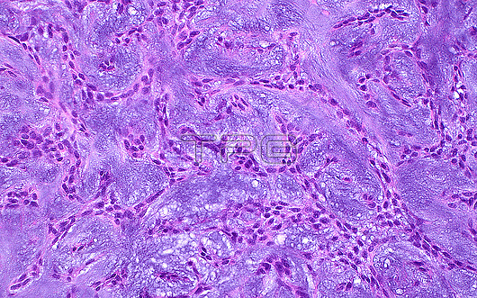
Light micrograph of a pleomorphic adenoma, a type of benign (non-cancerous) salivary gland tumour that is made up of epithelial and myoepithelial cells. The cells, which have oval round nuclei (small oval blue structures) are arranged in intersecting lines, within a so-called 'chondromyxoid stroma' (the dark blue-purple background in between the strands of cells). Haematoxylin and eosin stained tissue section. Magnification: x200 when printed at 10cm wide.
| px | px | dpi | = | cm | x | cm | = | MB |
Details
Creative#:
TOP29066908
Source:
達志影像
Authorization Type:
RM
Release Information:
須由TPG 完整授權
Model Release:
Not Available
Property Release:
Not Available
Right to Privacy:
No
Same folder images:
pathologymedicinepathologicalmedicalanatomicpathologysalivaryglandsalivaryglandtumourtumorparotidpleomorphicadenomasurgicalpathologyENTsurgeryheadandneckbiologyhistologyhistologicalhistopathologymicroscopylmlightmicrographslideHematoxylinHaematoxylinandeosinstainnobodyno-onehumanbodyanatomytissuecellsbenigntumourtumorchondromyxoidmatrixmyoepithelialcellspurplepurplebackgroundabstract
ENTHaematoxylinHematoxylinabstractadenomaanatomicanatomyandandbackgroundbenignbiologybodycellscellschondromyxoideosinglandglandheadhistologicalhistologyhistopathologyhumanlightlmmatrixmedicalmedicinemicrographmicroscopymyoepithelialneckno-onenobodyparotidpathologicalpathologypathologypathologypleomorphicpurplepurplesalivarysalivaryslidestainsurgerysurgicaltissuetumortumortumourtumour
 Loading