
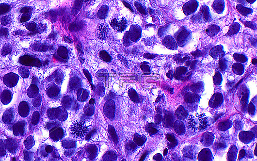
Light micrograph of mitotic figures. A few mitotic figures are seen amongst the many cancerous cells in this image of bladder cancer. The mitoses are dividing cells and are identified by their many small linear structures which are the condensed chromosomes of the cells. Haematoxylin and eosin stained tissue section. Magnification: 400x when printed at 10 cm.
| px | px | dpi | = | cm | x | cm | = | MB |
Details
Creative#:
TOP28634986
Source:
達志影像
Authorization Type:
RM
Release Information:
須由TPG 完整授權
Model Release:
n/a
Property Release:
n/a
Right to Privacy:
No
Same folder images:
pathologydiseasedisorderconditiondiagnosisdiagnosticmedicinehistologyhistologicalhistopathologydiseasedisorderconditiondiagnosisdiagnosticmicroscopylmmagnifiedimagelightmicrographslidehumanbodyanatomytissuecellsH&EstainHaematoxylinandeosinurinarybladderbladdercanceroncologymitosestumourgrowthdiseaseabnormal
H&EHaematoxylinabnormalanatomyandbladderbladderbodycancercellsconditionconditiondiagnosisdiagnosisdiagnosticdiagnosticdiseasediseasediseasedisorderdisordereosingrowthhistologicalhistologyhistopathologyhumanimagelightlmmagnifiedmedicinemicrographmicroscopymitosesoncologypathologyslidestaintissuetumoururinary
 Loading