
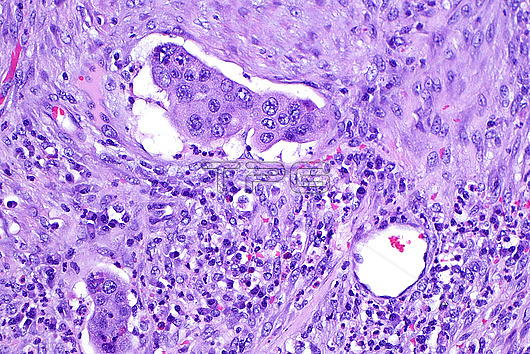
Light micrograph of a malignant tumour with lymphovascular invasion. The tumour cells are seen as a cluster within the white space of vessels at the top and bottom left of the image. Surrounding the vessels is connective tissue and small inflammatory cells Haematoxylin and eosin stained tissue section. Magnification: 200x when printed at 10 cm.
| px | px | dpi | = | cm | x | cm | = | MB |
Details
Creative#:
TOP28634983
Source:
達志影像
Authorization Type:
RM
Release Information:
須由TPG 完整授權
Model Release:
n/a
Property Release:
n/a
Right to Privacy:
No
Same folder images:
pathologydiseasedisorderconditiondiagnosisdiagnosticmedicineanatomicpathologydiseasedisorderconditiondiagnosisdiagnosticsurgicalpathologydiseasedisorderconditiondiagnosisdiagnostichistologyhistologicalhistopathologydiseasedisorderconditiondiagnosisdiagnosticmicroscopylmmagnifiedimagelightmicrographslidehumanbodyanatomytissuecellsH&EstainHaematoxylinandeosinlymphovascularinvasioncancerstagingcancerspreadmetastasisoncologydiseaseabnormal
H&EHaematoxylinabnormalanatomicanatomyandbodycancercancercellsconditionconditionconditionconditiondiagnosisdiagnosisdiagnosisdiagnosisdiagnosticdiagnosticdiagnosticdiagnosticdiseasediseasediseasediseasediseasedisorderdisorderdisorderdisordereosinhistologicalhistologyhistopathologyhumanimageinvasionlightlmlymphovascularmagnifiedmedicinemetastasismicrographmicroscopyoncologypathologypathologypathologyslidespreadstagingstainsurgicaltissue
 Loading