
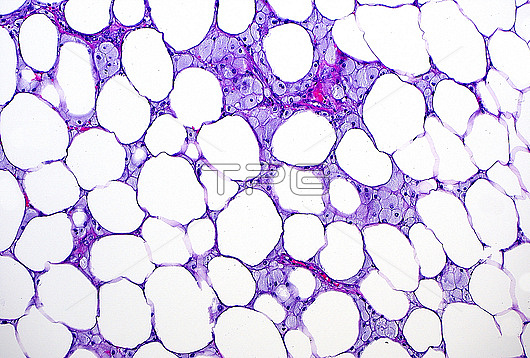
Light micrograph of fat necrosis. Dying fat cells (the white circular spaces) have no nuclei and are admixed with foamy histiocytes (the blue-gray cells in between). Haematoxylin and eosin stained tissue section. Magnification: 100x when printed at 10 cm.
| px | px | dpi | = | cm | x | cm | = | MB |
Details
Creative#:
TOP28634959
Source:
達志影像
Authorization Type:
RM
Release Information:
須由TPG 完整授權
Model Release:
n/a
Property Release:
n/a
Right to Privacy:
No
Same folder images:
 Loading