
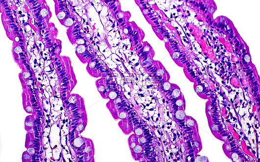
Light micrograph of the villi (finger-like structures) in the duodenum (first part of the small bowel). The inner portions of the villi have blood vessels (small bright pink areas) which take in the nutrients absorbed by the cells on the surface of the villi. Scattered goblet cells which are round and contain light blue mucin substance are also present on the surface of the villi. Haematoxylin and eosin stained tissue section. Magnification: 200x when printed at 10cm.
| px | px | dpi | = | cm | x | cm | = | MB |
Details
Creative#:
TOP28634934
Source:
達志影像
Authorization Type:
RM
Release Information:
須由TPG 完整授權
Model Release:
n/a
Property Release:
n/a
Right to Privacy:
No
Same folder images:
 Loading