
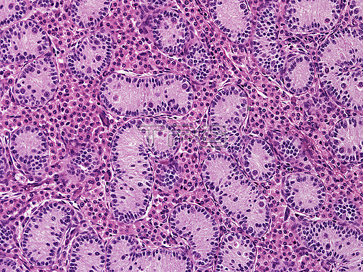
Foetal testis, light micrograph. The foetal testis shows seminiferous cords cut in various planes surrounded by an abundant population of Leydig cells that secrete androgens. The cords contain Sertoli cells arranged around the periphery with lesser numbers of round germ cells/spermatogonia in the central regions. In postnatal life spermatogonia activate to start spermatogenesis with the cords greatly enlarging in diameter and length to form seminiferous tubules. The foetal Leydig cell numbers regress after birth and a new population of adult-type Leydig cells develop. Paraffin section, haematoxylin and eosin stain. Magnification: x230 when width printed at 10cm.
| px | px | dpi | = | cm | x | cm | = | MB |
Details
Creative#:
TOP28610429
Source:
達志影像
Authorization Type:
RM
Release Information:
須由TPG 完整授權
Model Release:
n/a
Property Release:
n/a
Right to Privacy:
No
Same folder images:
 Loading