
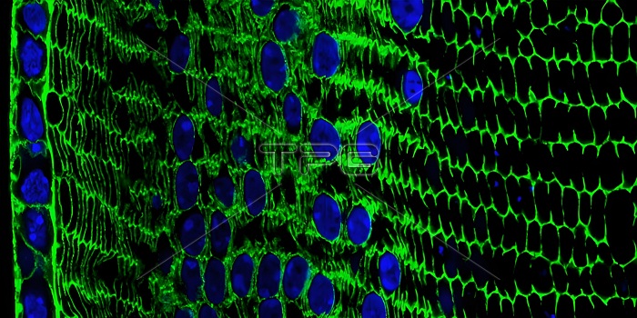
Here you can see the lens development process at work in a cross section of tissue from an adult mouse. In mice, as in humans, a single layer of rod-like epithelial cells (far left, blue/green) gives rise to specialized lens cells (middle, blue/green) throughout life. The new cells resemble their progenitor cells, with the nuclei (blue) and cytoskeletal protein actin (green). But soon these cells will produce large amounts of water-soluble proteins, called crystalline, to improve their transparency, while gradually breaking down their nuclei to eliminate light-scattering mass. What remains are fully differentiated, enucleated, non-replicating lens fibrous cells (right, green), which refract light onto the retina at the back of the eye.
| px | px | dpi | = | cm | x | cm | = | MB |
Details
Creative#:
TOP28024527
Source:
達志影像
Authorization Type:
RM
Release Information:
須由TPG 完整授權
Model Release:
N/A
Property Release:
N/A
Right to Privacy:
No
Same folder images:
 Loading