
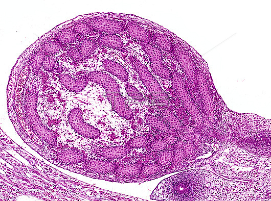
Light micrograph of foetal testis showing early development of seminiferous cords. Cords are full with germ cells and Sertoli cells and with later maturation and proliferation of the germ cells will form a central lumen to establish primitive seminiferous tubules. Seminiferous cords converge at one pole of the testis reflecting the postnatal anatomy where tubules will empty into the rete testis and ducts leading to the epididymis. Clusters of foetal Leydig cells are seen between some cords. Paraffin section, haematoxylin and eosin stain. Magnification: x60 when width printed at 10cm.
| px | px | dpi | = | cm | x | cm | = | MB |
Details
Creative#:
TOP27954919
Source:
達志影像
Authorization Type:
RM
Release Information:
須由TPG 完整授權
Model Release:
N/A
Property Release:
No
Right to Privacy:
No
Same folder images:
 Loading