
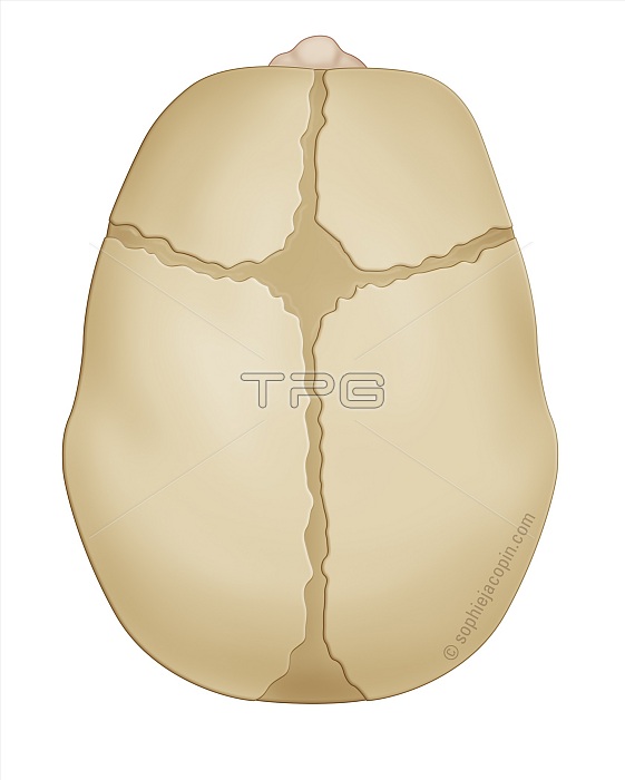
Bone structure at birth, bone development, cartilage structure, pediatrics. Infant skull in top view, fontanel, sutures, pediatrics. This anatomical illustration shows a newborn baby's skull viewed from above. The front of the skull is visible at the top of the drawing thanks to the nose represented. The skull is made up of bony plates, but also of fontanelles and sutures made of connective tissue. These structures give it great elasticity, allowing the baby to pass through the pelvic strait during birth. From top to bottom, we can distinguish the metopic suture, then the anterior fontanel. Coronal sutures are drawn on each side of the anterior fontanel. Then behind the anterior fontanel, it is the sagittal suture. This sagittal suture runs from the anterior fontanel to the posterior fontanel. Two lambdoid sutures, not shown here, start on each side of the posterior fontanel. In the baby, the bones of the skull are not united. This allows the skull to grow according to the rapid growth of the brain during the first years of life.
| px | px | dpi | = | cm | x | cm | = | MB |
Details
Creative#:
TOP27779150
Source:
達志影像
Authorization Type:
RM
Release Information:
須由TPG 完整授權
Model Release:
N/A
Property Release:
N/A
Right to Privacy:
No
Same folder images:
 Loading