
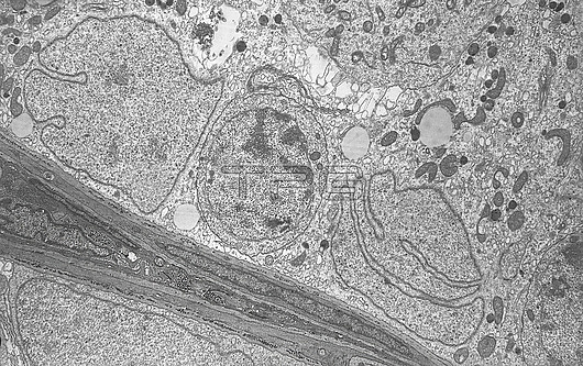
Transmission electron micrograph (TEM) of the ultrastructure of two Sertoli cell nuclei between which is an early (leptotene) primary spermatocyte. The basal lamina tissues of the seminiferous tubule is also shown consisting of several layers of myofibroblasts and connective tissue. Magnification: x6,000 when printed at a width of 10cm.
| px | px | dpi | = | cm | x | cm | = | MB |
Details
Creative#:
TOP27779071
Source:
達志影像
Authorization Type:
RM
Release Information:
須由TPG 完整授權
Model Release:
N/A
Property Release:
N/A
Right to Privacy:
No
Same folder images:
TestisseminiferousepitheliumspermatogenesisSertolicellspermatocyteprimaryspermatocytebasallaminamalemalereproductivetissuemalereproductivesystemultrastructurecellultrastructurecellbiologyelectronmicrographelectronmicroscopytransmissionelectronmicrographtemnobodyno-oneblackandwhitemonochromebiologicalcytologycytological
SertoliTestisandbasalbiologicalbiologyblackcellcellcellcytologicalcytologyelectronelectronelectronepitheliumlaminamalemalemalemicrographmicrographmicroscopymonochromeno-onenobodyprimaryreproductivereproductiveseminiferousspermatocytespermatocytespermatogenesissystemtemtissuetransmissionultrastructureultrastructurewhite
 Loading